. 2024 Jan 11;12(1):e0308023.
doi: 10.1128/spectrum.03080-23. Epub 2023 Nov 28.

Formulation and evaluation of atovaquone-loaded macrophage-derived exosomes against Toxoplasma gondii: in vitro and in vivo assessment

Affiliations

Formulation and evaluation of atovaquone-loaded macrophage-derived exosomes against Toxoplasma gondii: in vitro and in vivo assessment

Fatemeh Goudarzi et al. Microbiol Spectr. .

Abstract

This study is the first of its kind that suggests exosomes as a nano-carrier loaded with atovaquone (ATQ), which could be considered as a new strategy for improving the effectiveness of ATQ against acute and chronic phases of Toxoplasma gondii.

Keywords: atovaquone; drug delivery; exosome; in vitro; in vivo; toxoplasmosis.


Conflict of interest statement

The authors declare no conflict of interest.

Figures

Fig 1
Fig 1
Characterization of J774A.1 EXO. (A) Morphology of J774A.1 cells in two different conditions, FBS 10% and FBS 0%, under an inverted microscope. No significant morphological changes were detected. (B) Size distribution of J774A.1 EXO using dynamic light scattering (DLS) showed an average diameter of 84 nm. (C) TEM image of J774A.1 EXO showed the typical structure of exosomes. (D) SEM image of J774A.1 EXO showed spherical particles with diameters ranging between 38 and 128 nm. (E) Flow cytometry analysis revealed that isolated exosomes were positive for CD9 and CD63 exosomal markers.
Fig 2
Fig 2
Size distribution of EXO-ATQ using dynamic light scattering. The peak diameter was about 89 nm.
Fig 3
Fig 3
Cell viability assessment by MTT assays. Vero cells were treated with ATQ at different concentrations with or without exosomes for 48 h. S-ATQ and EXO-ATQ considerably reduced cell viability in a concentration-dependent manner with no significant differences (P > 0.05). Independent samples t test was used to make comparisons between groups.
Fig 4
Fig 4
(A) Infection rate and (B) the mean number of intracellular tachyzoites in the infected cells at different concentrations of compounds. EXO-ATQ significantly reduced the infection rate of T. gondii tachyzoites. The data represent three independent experiments and are presented as mean ± SD. One-way ANOVA was used to make comparisons between groups. *P < 0.05, **P < 0.01, ***P < 0.001, and ****P < 0.0001. (C) Microscopic images of tachyzoite-infected cells after 48 h of treatment with 240 µg/mL S-ATQ and EXO-ATQ compared to the untreated control group.
Fig 5
Fig 5
Toxicity studies including mice body, spleen, and liver weights in EXO-ATQ-treated and control groups. Results are presented as mean ± SD of four individual mice per group. No statistical differences were observed between the two groups (P > 0.05). Independent samples t test was used to make comparisons between groups.
Fig 6
Fig 6
(A) Serum levels of AST, ALT, and ALP of mice in the control and EXO-ATQ treatment groups. Values are expressed as mean  ±  SD of the mean. No significant difference between groups was observed (P > 0.05). Independent samples t test was used to make comparisons between groups. (B) Representative histological image of the liver and spleen of control and treated mice (magnification 1,000×). No histopathological abnormalities were observed in the treated mice compared with the control mice.
Fig 7
Fig 7
The anti-Toxoplasma efficiency of different compounds in female BALB/c mice infected with 1 × 10⁴ tachyzoites of T. gondii, RH strain, and virulence potential of post-treated tachyzoites. (A) The mice treated with EXO-ATQ had a significantly longer survival time than PBS-treated mice or those treated with S-ATQ, J774A.1 EXO, and DMSO. (B) The mean counts of peritoneum tachyzoites in mice treated with ATQ, especially EXO-ATQ, were significantly different from other groups. The data are presented as mean ± SD. ****P < 0.0001. One-way ANOVA was used to make comparisons. (C) Survival rates of the second group of mice inoculated with tachyzoites of T. gondii were obtained from the first group. The mice inoculated with tachyzoites obtained from EXO-ATQ-treated mice had a significantly longer survival time.
Fig 8
Fig 8
(A) The mean number and (B) size of tissue cysts in the brains of infected treated groups of mice compared to the infected non-treated control. The mean number and size of brain cysts were significantly reduced in the EXO-ATQ-treated group compared to the S-ATQ group. The data are presented as mean ± SD. One-way ANOVA was used to make comparisons between groups. *P < 0.05 and ****P < 0.0001.
Fig 9
Fig 9
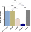
Mean expression ± SD of BAG1 in infected treated groups of mice and infected non-treated control. The real-time PCR analysis represented a significant reduction of BAG1 expression in the EXO-ATQ group compared to the other groups (P < 0.0001). One-way ANOVA was used to make comparisons between groups. ****P < 0.0001.
Fig 10
Fig 10
(A) The mean number and (B) size of tissue cysts in the brain of the second group of mice inoculated with bradyzoites obtained from the first group. Cyst burden in mice treated with EXO-ATQ was significantly different from those of S-ATQ-treated mice (P < 0.0001). One-way ANOVA was used to make comparisons between groups. ****P < 0.0001.

Similar articles

References

    1. Tartarelli I, Tinari A, Possenti A, Cherchi S, Falchi M, Dubey JP, Spano F. 2020. During host cell traversal and cell-to-cell passage, Toxoplasma gondii sporozoites inhabit the parasitophorous vacuole and posteriorly release dense granule protein-associated membranous trails. Int J Parasitol 50:1099–1115. doi:10.1016/j.ijpara.2020.06.012 - DOI - PubMed
    1. Ali-Heydari S, Keshavarz H, Shojaee S, Mohebali M. 2013. Diagnosis of antigenic markers of acute toxoplasmosis by IgG avidity immunoblotting. Parasite 20:18. doi:10.1051/parasite/2013017 - DOI - PMC - PubMed
    1. Rahbari AH, Keshavarz H, Shojaee S, Mohebali M, Rezaeian M. 2012. Igg avidity ELISA test for diagnosis of acute toxoplasmosis in humans. Korean J Parasitol 50:99–102. doi:10.3347/kjp.2012.50.2.99 - DOI - PMC - PubMed
    1. Teimouri A, Goudarzi F, Goudarzi K, Alimi R, Sahebi K, Foroozand H, Keshavarz H. 2022. Toxoplasma gondii infection in immunocompromised patients in Iran (2013-2022): a systematic review and meta-analysis. Iran J Parasitol 17:443–457. doi:10.18502/ijpa.v17i4.11271 - DOI - PMC - PubMed
    1. Zhou Z, Ortiz Lopez HIA, Pérez GE, Burgos LM, Farina JM, Saldarriaga C, Lopez-Santi R, Cotella JI, Pérez ALS, Baranchuk A. 2021. Toxoplasmosis and the heart. Curr Probl Cardiol 46:100741. doi:10.1016/j.cpcardiol.2020.100741 - DOI - PubMed

LinkOut - more resources