Review
. 2023 Nov 28;16(1):33.
doi: 10.1007/s40820-023-01258-4.

Nanoparticle Exsolution on Perovskite Oxides: Insights into Mechanism, Characteristics and Novel Strategies

Affiliations
Review

Nanoparticle Exsolution on Perovskite Oxides: Insights into Mechanism, Characteristics and Novel Strategies

Yo Han Kim et al. Nanomicro Lett. .

Abstract

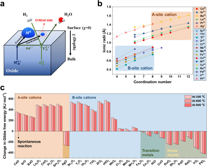
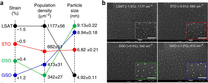
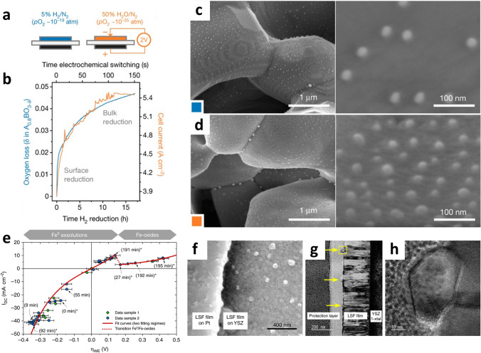
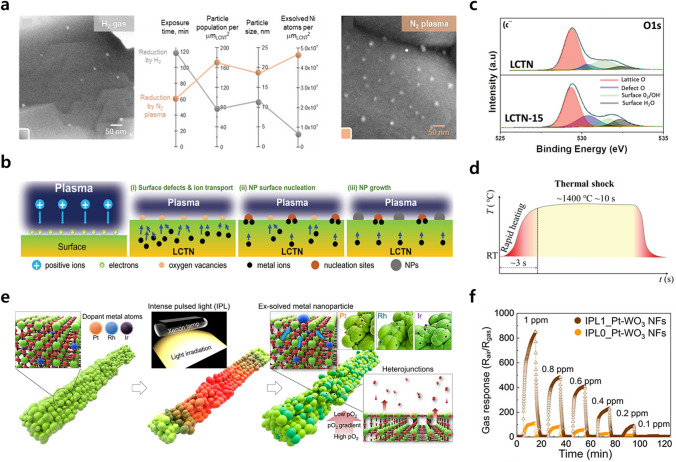
Supported nanoparticles have attracted considerable attention as a promising catalyst for achieving unique properties in numerous applications, including fuel cells, chemical conversion, and batteries. Nanocatalysts demonstrate high activity by expanding the number of active sites, but they also intensify deactivation issues, such as agglomeration and poisoning, simultaneously. Exsolution for bottom-up synthesis of supported nanoparticles has emerged as a breakthrough technique to overcome limitations associated with conventional nanomaterials. Nanoparticles are uniformly exsolved from perovskite oxide supports and socketed into the oxide support by a one-step reduction process. Their uniformity and stability, resulting from the socketed structure, play a crucial role in the development of novel nanocatalysts. Recently, tremendous research efforts have been dedicated to further controlling exsolution particles. To effectively address exsolution at a more precise level, understanding the underlying mechanism is essential. This review presents a comprehensive overview of the exsolution mechanism, with a focus on its driving force, processes, properties, and synergetic strategies, as well as new pathways for optimizing nanocatalysts in diverse applications.

Keywords: Catalyst; Exsolution; In situ growth; Mechanism; Perovskite oxide; Supported nanoparticle.


Conflict of interest statement

The authors declare no interest conflict. They have no known competing financial interests or personal relationships that could have appeared to influence the work reported in this paper.

Figures

Fig. 1
Fig. 1
a Schematic illustration of exsolution process under H2 gas reduction. b Ionic radius of metal cation candidates depending on coordination number. c Change in Gibbs free energy of oxide reduction reaction (1xMxOy+yxH2gM+yxH2Og) in H2 at 300, 600, and 900 °C in which the energy values are obtained using the HSC chemistry software
Fig. 2
Fig. 2
a Exsolved nuclei on the oxide surface with a wetting angle. b Cross-sectional STEM image of exsolved Ni nanoparticles on the La0.2Sr0.7Ni0.1Ti0.9O3δ after heating treatment inside STEM at 700 °C. c Schematic illustration of preferential Ni exsolution near anti-phase boundaries. Time-dependent evolution of size of d Ni particle and e crystalline Ni. Reprinted permission from Ref. [78]. f Scenarios of exsolution process in early stage. Reprinted permission from Ref. [79]
Fig. 3
Fig. 3
a TEM image of an exsolved Ni nanoparticle on the perovskite oxide. Surface morphologies of b Ni-exsolved perovskite oxide and c a sample etched by HNO3. d 3D AFM image of a socket after etching. Reprinted permission from Ref. [61]. e Environmental TEM analysis of exsolution particles with different reduction time (t) in H2. f Schematic illustration of nucleation and socketing during particle growth in the exsolution process. Reprinted permission from Ref. [59]. g, h In situ observation of reactive wetting during growth. Reprinted permission from Ref. [78]. i Plot for Gibbs free energies of supported particles with different embedment. Reprinted permission from Ref. [87]
Fig. 4
Fig. 4
a Particle growth with isotropic relation between height and width. b The number of exsolved Ni atoms. Reprinted permission from Ref. [59]. c Radius of exsolved Co particle as function of time with fitted lines of analytic models. Reprinted permission from Ref. [88]. TEM images of exsolved material with EDS results of d points and e linear scanning. f Corresponding schematics of particle distribution. Reprinted permission from Ref. [90]. g Concentration of Fe metal on the surface of thin films with different thickness as a function of the elapsed time. h Schematic illustration of exsolution behavior limited by surface reduction. Reprinted permission from Ref. [60]
Fig. 5
Fig. 5
Morphologies of reported metallic exsolution particles with different shapes. a Spherical Ni. Reprinted permission from Ref. [90]. b Cubic Ni. Reprinted permission from Ref. [59]. c Triangular Ni. d Pyramidal Ni. Reprinted permission from Ref. [87]. e, f Cone-shaped CoFe alloy. Reprinted permission from Ref. [42]. g Triangular Pt. Reprinted permission from Ref. [65]
Fig. 6
Fig. 6
a Cross-sectional morphologies of exsolved Ni particles with different size. b Plot of shape factor as function of interfacial energy. Reprinted permission from Ref. [87]
Fig. 7
Fig. 7
a Theoretical and experimental exsolution results on different reduction time and PO2 condition. Reprinted permission from Ref. [72]. b Exsolution characteristics under different reduction temperatures. Reprinted permission from Ref. [34]. c Morphologies of exsolved Co particles in the STF thin film after reduction at 450–550 °C. d Corresponding particle tendency regarding size, population density, and the exsolution extent. Reprinted permission from Ref. [103]
Fig. 8
Fig. 8
Effect of stoichiometry on exsolution perovskite design. a Thermogravimetric analysis (TGA), b the extent of oxygen vacancy formation, and c H2-TPR of the LSC, 73LSCNi-15, and 63LSCNi-15. Reprinted permission from Ref. [107]. d Surface exsolution of Ni nanoparticles on the A-site deficient perovskite. e Surface exsolution of Ni nanoparticles on the perovskites without defects. Reprinted permission from Ref. [38]
Fig. 9
Fig. 9
a Nanoparticle exsolution on different surface termination. b AFM image and atomic scale perovskite model with different surface orientation. Reprinted permission from Ref. [61]. c Exsolution of nanometal particles on epitaxially prepared perovskite thin film. Reprinted permission from Ref. [71]. d Calculated Ni segregation energy on different surface orientation. Reprinted permission from Ref. [67]
Fig. 10
Fig. 10
a Size and distribution of exsolved nanoparticles as a function of surface strain. b SEM images of exsolved nanoparticles prepared on diverse strain-induced perovskite thin films. Reprinted permission from Ref. [115]
Fig. 11
Fig. 11
Thermal stability characteristics of exsolved nanocatalysts. a Comparison between infiltrated and exsolved Cu nanoparticles in dry H2 at 600 °C as a function of reduction time. (Cu particles are highlighted in blue in SEM image). Reprinted permission from Ref. [21]. b Comparison between exsolved and impregnated Ru nanoparticles after the ammonia synthesis test. Reprinted permission from Ref. [81]
Fig. 12
Fig. 12
Remarkable anti-coking trait of exsolved catalysts. a SEM images of infiltrated and exsolved Ni particles after coking test in 20% CH4/H2, at 800 °C for 4 h. (scale bars, 0.5 mm (overview); 100 nm (detail)). Reprinted permission from Ref. [61]. b Schematic of possible carbon fiber growth mechanisms. c Activity of shape-controlled Ni exsolution and reference catalysts in dry reforming of methane at 700 °C (GHSV = 30,000 mL g−1 h−1). Reprinted permission from Ref. [87]
Fig. 13
Fig. 13
Regenerative capability of exsolved particles. a Schematic illustration of self-regeneration mechanism. Reprinted permission from Ref. [55]. b The redox reversibility and stable long-term performance of a perovskite anode material, La0.3Sr0.7Cr0.3Fe0.6Co0.1O3 − δ with exsolved Co–Fe nanoparticles. Reprinted permission from Ref. [121]
Fig. 14
Fig. 14
Electrochemical switching to trigger exsolution on the perovskite oxide. a Reduction conditions and b reduction tendencies of fuel electrodes using gas reduction and electrochemical switching methods. Morphologies of electrode surface after c traditional H2 reduction for 20 h and d electrochemical switching process (2 V, 150 s). Reprinted permission from Ref. [1]. e Plot of the area-normalized current (IDC) versus overpotential dropping at the LSF thin film working electrode. f–h Electron microscopic images of exsolved Fe nanoparticles on the LSF thin film after 60 h under reduction conditions. Reprinted permission from Ref. [123]
Fig. 15
Fig. 15
a SEM and characteristics analysis of the Ni nanoparticles grown on the La0.43Ca0.37Ni0.06Ti0.94O3 − d perovskite surface after exposure to H2 gas for 2 h (gray) and N2 plasma for 1 h (orange) at 650 °C. Reprinted permission from Ref. [86]. b Schematic representation of plasma-driven exsolution of Ni nanoparticles indicating various phenomena occurring during the process. c XPS results of O 1s of LCNT before (LCNT) and after (LCNT-15) Ar plasma treatment. Reprinted permission from Ref. [125]. d Temperature profile for the thermal shock technique. Reprinted permission from Ref. [126]. e Schematic diagram of exsolved noble metals decoration on WO3 NFs via IPL-MP treatment with a mechanism of heterojunctions and exsolution formation. f Hydrogen sulfide (H2S) gas sensing results from 1 to 100 ppb concentration of IPL exsolved Pt–WO3 NFs. Reprinted permission from Ref. [127]
Fig. 16
Fig. 16
Exsolution and phase transition properties in perovskite systems. a DFT calculated profiles of Gibbs free energy for oxygen vacancy formation from the surface AO (A-site) and BO2 (B-site) networks. b Total phase transition energy from single to RP perovskite. c Oxygen vacancy formation and co-segregation energies as function of Sr doping ratio. Reprinted permission from Ref. [137]. d SE-STEM images of SFRuM after 4.th repeated phase transition and population and size of exsolved nanoparticles as a function of redox number. e Segregation energies of Fe and Ru in diverse conditions. f Relative energy of the slabs as a function of Ru position. Reprinted permission from Ref. [89]
Fig. 17
Fig. 17
Bimetal exsolution strategies for triggering nanoparticles formation. a Schematic illustration and surface morphologies of Co exsolution with topotactic ionic exchange method. Reprinted permission from Ref. [135]. b Schematic illustration of seeded effect for Cu–Fe nanoparticle exsolution. Reprinted permission from Ref. [64]

References

    1. J.H. Myung, D. Neagu, D.N. Miller, J.T.S. Irvine, Switching on electrocatalytic activity in solid oxide cells. Nature 537, 528–531 (2016). 10.1038/nature19090 - PubMed
    1. P. Zhou, I.A. Navid, Y. Ma, Y. Xiao, P. Wang et al., Solar-to-hydrogen efficiency of more than 9% in photocatalytic water splitting. Nature 613, 66–70 (2023). 10.1038/s41586-022-05399-1 - PubMed
    1. X. Li, L. Dai, Z. He, W. Meng, Y. Li et al., In situ exsolution of PdO nanoparticles from non-stoichiometric LaFePd0.05O3+δ electrode for impedancemetric NO2 sensor. Sens. Actuators B Chem. 298, 126827 (2019). 10.1016/j.snb.2019.126827
    1. S.H. Kim, H. Jeong, B. Sharma, J.H. Myung, In situ exsolution catalyst: An innovative approach to develop highly selective and sensitive gas sensors. ACS Appl. Mater. Interfaces 14, 18275–18282 (2022). 10.1021/acsami.1c22701 - PubMed
    1. M. Akri, S. Zhao, X. Li, K. Zang, A.F. Lee et al., Atomically dispersed nickel as coke-resistant active sites for methane dry reforming. Nat. Commun. 10, 5181 (2019). 10.1038/s41467-019-12843-w - PMC - PubMed

LinkOut - more resources