Review
. 2024 Apr;54(4):490-504.
doi: 10.1007/s00247-023-05815-2. Epub 2023 Nov 28.

A clinical perspective on imaging in juvenile idiopathic arthritis

Affiliations
Review

A clinical perspective on imaging in juvenile idiopathic arthritis

Maria Tarsia et al. Pediatr Radiol. 2024 Apr.

Abstract

In recent years, imaging has become increasingly important to confirm diagnosis, monitor disease activity, and predict disease course and outcome in children with juvenile idiopathic arthritis (JIA). Over the past few decades, great efforts have been made to improve the quality of diagnostic imaging and to reach a consensus on which methods and scoring systems to use. However, there are still some critical issues, and the diagnosis, course, and management of JIA are closely related to clinical assessment. This review discusses the main indications for conventional radiography (XR), musculoskeletal ultrasound (US), and magnetic resonance imaging (MRI), while trying to maintain a clinical perspective. The diagnostic-therapeutic timing at which one or the other method should be used, depending on the disease/patient phenotype, will be assessed, considering the main advantages and disadvantages of each imaging modality according to the currently available literature. Some brief clinical case scenarios on the most frequently and severely involved joints in JIA are also presented.

Keywords: Children; Conventional radiography; Imaging; Juvenile idiopathic arthritis; Magnetic resonance imaging; Ultrasound.


Conflict of interest statement

None

Figures

Fig. 1
Fig. 1
Anteroposterior radiograph of both hands in a 13.5-year-old boy with enthesitis-related (HLA B27 positive) juvenile idiopathic arthritis: general mild osteoporosis of the right hand (long arrow), soft tissue edema around the carpal bones on the right (short arrow), narrowed carpometacarpal joint spaces on the right (long thin arrow), discrepancy of the size of the carpal bones due to accelerated growth on the right (short thin arrow), suspicious erosions at the base of the second to fourth metacarpals and at os trapezoideum and os capitatum (arrowheads). Normal structure and morphology of the bones of the left hand
Fig. 2
Fig. 2
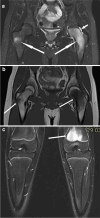
Magnetic resonance images in a 15-year-old boy with chronic recurrent multifocal osteomyelitis. a, b Coronal T2 turbo inversion recovery magnitude (TIRM) (a) and T1 (b) images of the hips show bone edema in the metaphysis and epiphysis of the left femur and at the base of the right greater trochanter (long arrows). There is a small reactive effusion in the left hip joint (short arrow in a). c Coronal T2 TIRM image shows bone edema in the distal metaphysis of the left femur (arrow)
Fig. 3
Fig. 3
Anteroposterior radiographs of both hands in a 19-year-old young woman with a long history of aggressive seronegative polyarticular juvenile idiopathic arthritis, onset at the age of 7 years and with poor adherence to treatment. Images show mild bilateral periarticular osteoporosis (long arrows), growth arrest lines in the radial metaphysis and bone-in-bone appearance of carpal bones (short arrows), joint space narrowing of carpo-metacarpal joints and intercarpal bones, more pronounced on the right (arrowheads), and mild soft tissue edema around the right ulna (thin arrow)
Fig. 4
Fig. 4
Power Doppler ultrasound of left talonavicular joint in sagittal projection in a 19-year-old young woman with a long history of aggressive seronegative polyarticular juvenile idiopathic arthritis, onset at the age of 7 years and with poor adherence to treatment (same patient as in Fig. 3) shows thickened and chronically altered synovium without a joint effusion or significant hyperemia (arrow)
Fig. 5
Fig. 5
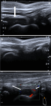
Ankle ultrasound of a 2-year-old girl with juvenile idiopathic arthritis (HLA B27 negative, anti-nuclear antibodies positive). a, b Sagittal projection of right (a) and left (b) ankle with effusion in the right side (arrow); (c) Sagittal power Doppler study of right ankle with effusion (long arrow), and thickened and hyperemic synovium (short arrow)—signs of synovitis
Fig. 6
Fig. 6
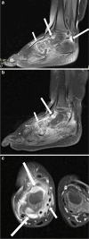
Magnetic resonance imaging of the right ankle in a 2-year-old girl with juvenile idiopathic arthritis (HLA B27 negative, anti-nuclear antibodies positive, same patient as in Fig. 5). a Sagittal proton-density-weighted fat suppressed image shows marked synovial proliferation and effusion of the anterior and posterior recesses of the ankle joint (long arrows) and around the extensor tendons (short arrow)—signs of synovitis and tenosynovitis. b Sagittal postcontrast T1-weighted fat-suppressed sequence shows marked synovial proliferation and enhancement of the anterior recess of the ankle joint (long arrow) and around the tarsal bones (short arrow)—signs of active synovitis. c Axial postcontrast T1-weighted fat-suppressed sequence shows marked synovial proliferation and enhancement of the anterior and posterior recesses of the ankle joint (long arrows) and around the flexor tendons (short arrow)—signs of active synovitis and tenosynovitis
Fig. 7
Fig. 7
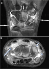
Magnetic resonance imaging of the right wrist in a 14-year-old boy with juvenile idiopathic arthritis with enthesitis (HLA B27 positive). Coronal (a) and axial (b) proton-density-weighted fat-suppressed images show marked synovial proliferation (long arrows), reactive edema of the carpal bones (short arrows), and a small joint effusion (arrowhead in b)—signs of synovitis
Fig. 8
Fig. 8
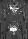
Coronal magnetic resonance imaging of the hips in a 15-year-old girl with psoriatic arthritis (HLA B27 negative, ANA positive). T2 turbo inversion recovery image shows an effusion in the left hip joint (arrow); bone and cartilage are normal in appearance. b Postcontrast T1-weighted fat-suppressed image shows synovial enhancement on the left (arrow)—a sign of active synovitis
Fig. 9
Fig. 9
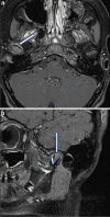
Postcontrast T1-weighted fat-suppressed magnetic resonance images of the temporomandibular joints in a 14-year-old girl with oligoarticular juvenile idiopathic arthritis (ANA positive). a Axial image shows synovial thickening and enhancement on the right (arrow)—a sign of active synovitis. b Sagittal image shows a flattened mandibular head (long arrow) and synovial thickening and enhancement on the right (short arrow)—signs of chronic bone changes and active synovitis

Similar articles

Cited by

References

    1. Thierry S, Fautrel B, Lemelle I, Guillemin F. Prevalence and incidence of juvenile idiopathic arthritis: a systematic review. Joint Bone Spine. 2014;81:112–117. - PubMed
    1. Brewer EJ, Jr, Bass J, Baum J, et al. Current proposed revision of JRA Criteria. JRA Criteria Subcommittee of the Diagnostic and Therapeutic Criteria Committee of the American Rheumatism Section of The Arthritis Foundation. Arthritis Rheum. 1977;20:195–199. - PubMed
    1. Carol HA, Chauvin NA, Weiss PF. Imaging in pediatric spondyloarthritis. Curr Opin Rheumatol. 2023;35:226–234. - PMC - PubMed
    1. Malattia C, Tzaribachev N, van den Berg JM, Magni-Manzoni S. Juvenile idiopathic arthritis - the role of imaging from a rheumatologist’s perspective. Pediatr Radiol. 2018;48:785–791. - PubMed
    1. Navallas M, Tolend M, Otobo TM, et al. Developing standards for MRI evaluation of joints in children with juvenile idiopathic arthritis utilizing the temporomandibular joint as a model. Jpn J Radiol (in press) 2023 doi: 10.1007/s11604-023-01479-y. - DOI - PubMed

Publication types