. 2023 Nov 1;7(11):771-787.
doi: 10.4049/immunohorizons.2300094.

PD-1 Impairs CD8+ T Cell Granzyme B Production in Aged Mice during Acute Viral Respiratory Infection

Affiliations

PD-1 Impairs CD8+ T Cell Granzyme B Production in Aged Mice during Acute Viral Respiratory Infection

Olivia B Parks et al. Immunohorizons. .

Abstract

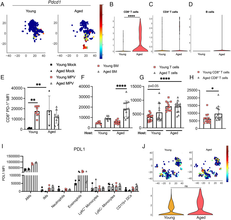
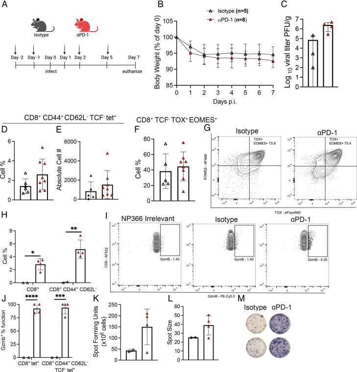
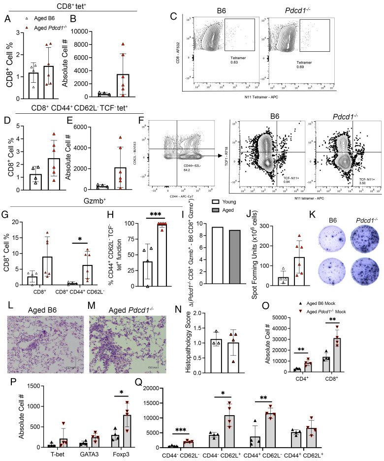
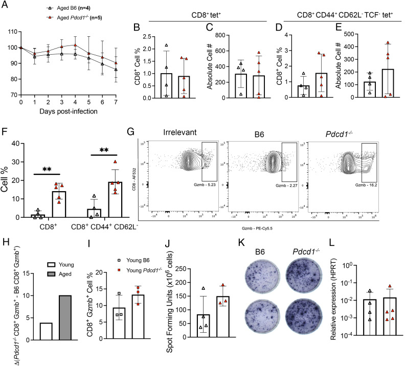
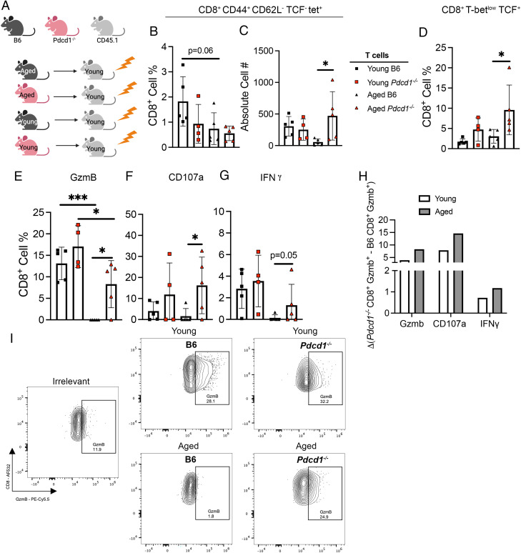
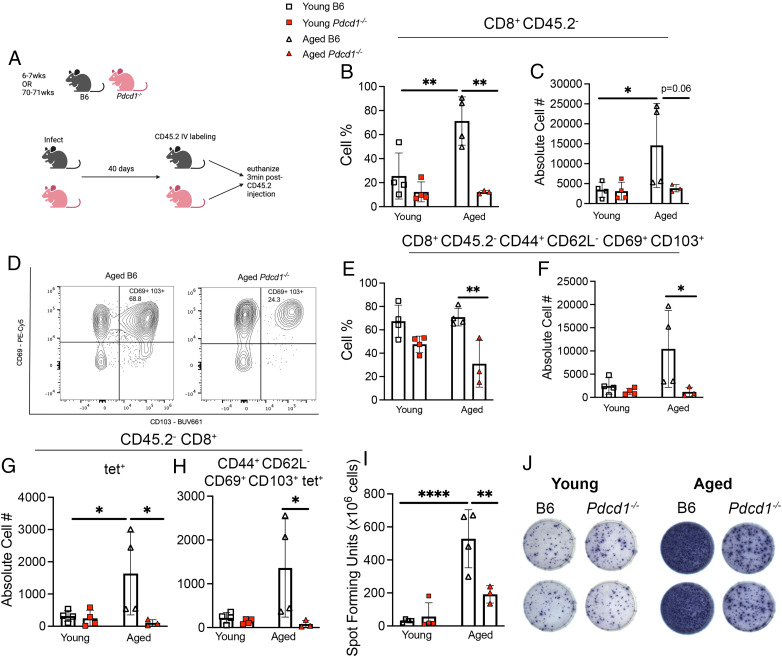
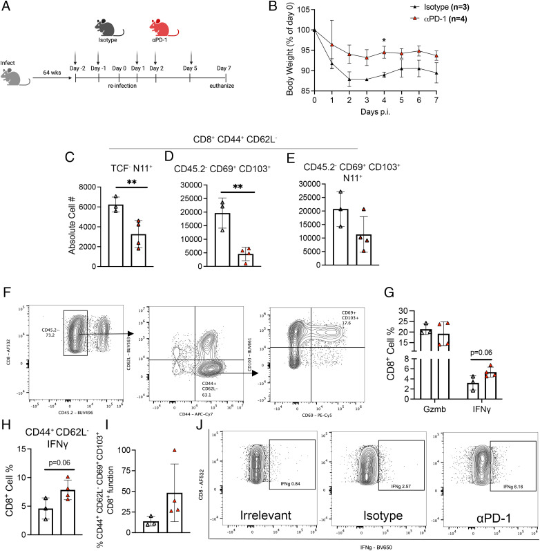
CD8+ T cell dysfunction contributes to severe respiratory viral infection outcomes in older adults. CD8+ T cells are the primary cell type responsible for viral clearance. With increasing age, CD8+ T cell function declines in conjunction with an accumulation of cytotoxic tissue-resident memory (TRM) CD8+ T cells. We sought to elucidate the role of PD-1 signaling on aged CD8+ T cell function and accumulation of CD8+ TRM cells during acute viral respiratory tract infection, given the importance of PD-1 regulating CD8+ T cells during acute and chronic infections. PD-1 blockade or genetic ablation in aged mice yielded improved CD8+ T cell granzyme B production comparable to that in young mice during human metapneumovirus and influenza viral infections. Syngeneic transplant and adoptive transfer strategies revealed that improved granzyme B production in aged Pdcd1-/- CD8+ T cells was primarily cell intrinsic because aged wild-type CD8+ T cells did not have increased granzyme B production when transplanted into a young host. PD-1 signaling promoted accumulation of cytotoxic CD8+ TRM cells in aged mice. PD-1 blockade of aged mice during rechallenge infection resulted in improved clinical outcomes that paralleled reduced accumulation of CD8+ TRM cells. These findings suggest that PD-1 signaling impaired CD8+ T cell granzyme B production and contributed to CD8+ TRM cell accumulation in the aged lung. These findings have implications for future research investigating PD-1 checkpoint inhibitors as a potential therapeutic option for elderly patients with severe respiratory viral infections.


Conflict of interest statement

The authors have no financial conflicts of interest.

Figures

FIGURE 1.
FIGURE 1.
Aged CD8+ T cells upregulate PD-1 at baseline and during HMPV infection. (A) Heat map of Pdcd1 on lung CD8+ T cells from uninfected, naive aged and young mice. (BD) Violin plot of Pdcd1−/− expression from uninfected, naive aged and young mice on lung CD8+ T, CD4+ T, and B cells, respectively. (E) Mean fluorescence intensity (MFI) of PD-1 expression on CD8+ T cells in young and aged mock-infected or infected mice. (FH) MFI of PD-1 expression on lung CD8+ T cells in day 7 p.i. in (F) aged/young mixed BM chimera model, (G) syngeneic transplant of bulk aged or young T cells along with young B cells and Rag1−/− BM, and (H) adoptive transfer of aged or young CD8+ T cells into young Rag1−/− recipients. (I) PDL1 MFI expression on lung innate immune cells at day 1 p.i. (J) Heat map of PD-L1 (i.e., CD274) expression in lung myeloid cell cluster in uninfected, naive young and aged mice (top) and violin plot (bottom). Each data point represents one individual mouse. Data in (E) represent three experimental replicates, one to three mouse/group. Data in (F) represent two experimental replicates, three to seven mice/group. Data in (G) represent four experimental replicates, three to five mice/group. Data in (H) represent two experimental replicates, six or seven mice/group. Data in (I) represent one experimental replicate, three mice/group. *p < 0.05, **p < 0.01, ****p < 0.0001, unpaired t test or one-way ANOVA. AM, alveolar macrophages; DC, dendritic cells; IM, interstitial macrophages; MPV, metapneumovirus.
FIGURE 2.
FIGURE 2.
Proportion of granzyme B–expressing CD8+ T cells increases after PD-1 blockade. (A) Aged mice were treated with 200 g/200 l PD-1 or rat isotype control Ab via i.p. injection 2 d prior to infection and days 1, 2, and 5 p.i. (B) Weight loss during infection. (C) Viral titer in PFU/g between aged mice treated with isotype control of PD-1 blockade. (D and E) Cell percentage and absolute cell number of lung CD44+ CD62L TCF tet+ CD8+ T cells. (F) TCF TOX+ EOMES+ lung CD8+ T cells in aged mice treated with isotype or PD-1 blockade. (G) Representative flow plots of TOX and EOMES staining. (H) Bulk lung CD8+ and CD44+ CD62L Gzmb+ cell percentage. (I) Representative flow plots of Gzmb staining on lung CD8+ CD44+ CD62L T cells. (J) GzmB percentage function of lung CD8+ tet+ and CD8+ CD44+ CD62L TCF tet+ in both groups. (K) Spot number from ex vivo peptide stimulation IFN-γ ELISPOT of lung lymphocytes from aged isotype- or PD-1 blockade–treated mice and (L) spot size. (M) Representative images of ELISPOT wells. Absolute cell number calculation by BioLegend Precision Counting Beads. Each data point represents one individual mouse. Data in (B)–(G) represent three experimental replicates with two to four mice/group. Data in (H)–(J) represent two experimental replicates with two or three mice/group. Data in (K)–(M) represent one experimental replicate with two to four mice/group. *p < 0.05, **p < 0.01, ***p < 0.001, ****p < 0.0001.
FIGURE 3.
FIGURE 3.
Aged Pdcd1−/− mice have improved CD8+ T cell granzyme B production during HMPV infection. (A and B) Cell percentage (left) and absolute cell number (right) of lung CD8+ tet+ T cells in B6 and Pdcd1−/− mice at day 7 p.i. (C) Representative flow plots of tetramer staining. (D and E) Cell percentage (left) and absolute cell number (right) of lung CD8+ CD44+ CD62L TCF tet+ cells in aged B6 and Pdcd1−/− mice at day 7 p.i. (F) Representative flow plots of TCF tetramer staining. (G) Granzyme B+ bulk CD8+ and CD44+ CD62L CD8+ T cells. (H) Percentage function of granzyme B+ CD44+ CD62L TCF tet+ lung CD8+ T cells. (I) Δ Change of Pdcd1−/− CD8+ Gzmb+ T cells minus B6 CD8+ Gzmb+ T cells in young and aged HMPV-infected mice at day 7 p.i. (J) ELISPOT ex vivo peptide stimulation IFN-γ production from B6 or Pdcd1−/− lung lymphocytes. (K) Representative ELISPOT well images. (L and M) H&E images of aged B6 and Pdcd1−/− lungs on day 7 p.i. (N) Histopathological scoring from both groups of mice. (O) Absolute cell number of CD4+ and CD8+ T lymphocytes, (P) T-bet, GATA3, Foxp3+ CD4+ T lymphocytes, and (Q) CD44 and CD62L expression on CD8+ T lymphocytes in aged B6 mice or Pdcd1−/− mock-infected mice. Absolute cell number calculation by BioLegend Precision Counting Beads. The histopathological score is the average score per section field by a group-blinded experienced lung pathologist: 0, no inflammation; 1, <25% inflammation; 2, 25–50% inflammation; 3, 50–75% inflammation; and 4, >75% inflammation. Each data point represents one individual mouse. Data in (A)–(K) represent two experimental replicates with two or three mice/group. Data in (L) and (M) represent one experimental replicate with three or four mice/group. *p < 0.05, **p < 0.01, ***p < 0.001, unpaired t test or one-way ANOVA.
FIGURE 4.
FIGURE 4.
Aged Pdcd1−/− influenza-infected mice also exhibited improved CD8+ T cell granzyme B production. (A) Weight loss of aged B6 and Pdcd1−/− mice during influenza PR8 infection. (B and C) Bulk lung CD8+ tet+ cell percentage and absolute cell number. (D and E) Terminally differentiated lung CD8+ CD44+ CD62L TCF tet+ cell percentage and absolute cell number. (F) Bulk lung CD8+ T cell and CD8+ CD44+ CD62L granzyme B production. (G) Representative flow plots of granzyme B staining. (H) Δ Change of Pdcd1−/− CD8+ Gzmb+ T cells minus B6 CD8+ Gzmb+ in lungs of young and aged PR8-infected mice. (I) Lung CD8+ granzyme B+ cell percentage in young B6 or Pdcd1−/− mice. (J) Spot-forming units from IFN-γ ELISPOT ex vivo class I peptide stimulation from aged B6 and Pdcd1−/− lung lymphocytes. (K) Representative images of wells from IFN-γ ELISPOT. (L) Quantitative RT-PCR relative expression to housekeeping gene (HPRT) or influenza A transcripts in lung homogenate. Absolute cell number calculation by BioLegend Precision Counting Beads. Each data point represents one individual mouse. Data (A)–(G) and (I) represent two experimental replicates with two or three mice/group. Data in (H) represent one experimental replicate with three mice per group. Data in (J)–(L) represent one experimental replicate with three or four mice/group. **p < 0.05, unpaired t test or two-way ANOVA.
FIGURE 5.
FIGURE 5.
Aged Pdcd1−/− CD8+ T cells transplanted into young mice had increased granzyme B production. (A) Experimental schematic. Aged or young B6 or Pdcd1−/− T cells, young B6 CD19+ B cells, and young Rag1−/− BM were transplanted into lethally irradiated CD45.1 young recipients. At 6 wk after transplant, mice were infected with HMPV, and CD8+ T cell response was assessed on day 7 p.i. (B and C) TCF tet+ lung CD8+ cell percentage and absolute cell number in all four groups. (D) Cell percentage of stem cell–like T-betlow TCF+ lung CD8+ T cells. (EG) Lung CD8+ T cells positive for granzyme B, CD107a, and IFN-γ, respectively. (H) Δ Change of donor Pdcd1−/− CD8+ Gzmb+ T cells minus B6 CD8+ Gzmb+ T cells in young HMPV-infected recipient. (I) Representative flow plots of granzyme B staining. Each data point represents one individual mouse. Data represent one experimental replicate with four or five mice/group. *p < 0.05, ***p < 0.001, one-way ANOVA.
FIGURE 6.
FIGURE 6.
Aged Pdcd1−/− had fewer TRM cells at 40 d p.i. (A) Experimental schematic. (B and C) CD8+ CD45.2 cell percentage and absolute cell number in lungs of young and aged B6 and Pdcd1−/− mice. (D) Representative flow plots of CD69 and CD103 staining. (E and F) Cell percentage and absolute cell number of CD8+ CD45.2 CD44+ CD62L CD69+ CD103+ in all four groups. (G and H) Absolute cell number of bulk lung CD45.2 CD8+ tet+ and memory CD44+ CD62L CD69+ CD103+ T cells in all four groups. (I) Spot-forming units from IFN-γ ELISPOT ex vivo class I peptide stimulation from lung lymphocytes. (J) Representative images from IFN-γ ELISPOT wells. Absolute cell number calculated using BioLegend Precision Counting Beads. Each data point represents one individual mouse. Data represent one experimental replicate with three or four mice/group. *p < 0.05, **p < 0.01, ****p < 0.0001, one-way ANOVA.
FIGURE 7.
FIGURE 7.
PD-1 blockade improves weight loss and memory CD8+ T cell function during reinfection of aged mice. (A) Experimental schematic. (B) Weight loss after rechallenge. (CE) Absolute cell number of lung CD8+ CD44+ CD62L. (F) Representative flow plots of CD69 and CD103 staining. (G) Lung CD8+ Gzmb+ and IFN-γ+ cell percentage in both groups. (H) CD8+ CD44+ CD62L IFN-γ+ cell percentage in lung. (I) Memory CD8+ T cell IFN-γ percentage function in lung. (J) Representative flow plots of IFN-γ staining. Absolute cell number calculation by BioLegend Precision Counting Beads. Each data point represents one individual mouse. Data represent one experimental replicate with three or four mice/group. *p < 0.05, **p < 0.01, unpaired t test or two-way ANOVA.

Similar articles

Cited by

References

    1. Shafagati, N., Williams J.. 2018. Human metapneumovirus - what we know now. F1000 Res. 7: 135. - PMC - PubMed
    1. Falsey, A. R., Erdman D., Anderson L. J., Walsh E. E.. 2003. Human metapneumovirus infections in young and elderly adults. J. Infect. Dis. 187: 785–790. - PubMed
    1. Hasvold, J., Sjoding M., Pohl K., Cooke C., Hyzy R. C.. 2016. The role of human metapneumovirus in the critically ill adult patient. J. Crit. Care 31: 233–237. - PMC - PubMed
    1. van den Hoogen, B. G., Osterhaus D. M., Fouchier R. A.. 2004. Clinical impact and diagnosis of human metapneumovirus infection. Pediatr. Infect. Dis. J. 23(1 Suppl): S25–S32. - PubMed
    1. Widmer, K., Zhu Y., Williams J. V., Griffin M. R., Edwards K. M., Talbot H. K.. 2012. Rates of hospitalizations for respiratory syncytial virus, human metapneumovirus, and influenza virus in older adults. J. Infect. Dis. 206: 56–62. - PMC - PubMed

Publication types

Substances