. 2024 Jan 16;134(2):e166134.
doi: 10.1172/JCI166134.

Age-related loss of Notch3 underlies brain vascular contractility deficiencies, glymphatic dysfunction, and neurodegeneration in mice

Affiliations

Age-related loss of Notch3 underlies brain vascular contractility deficiencies, glymphatic dysfunction, and neurodegeneration in mice

Milagros C Romay et al. J Clin Invest. .

Abstract

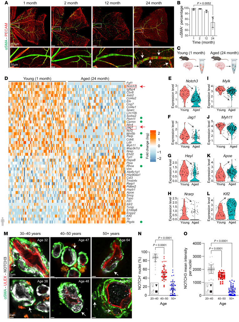
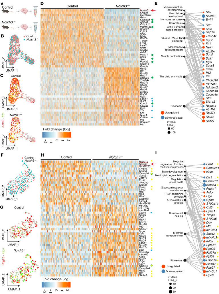
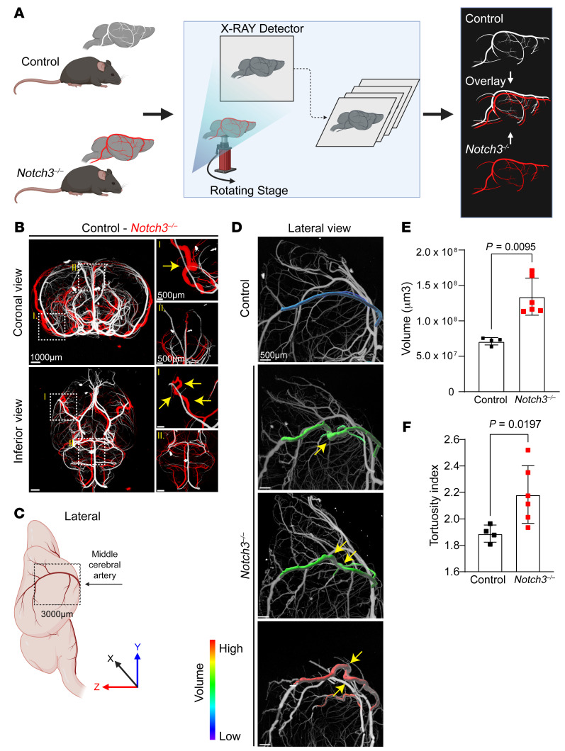
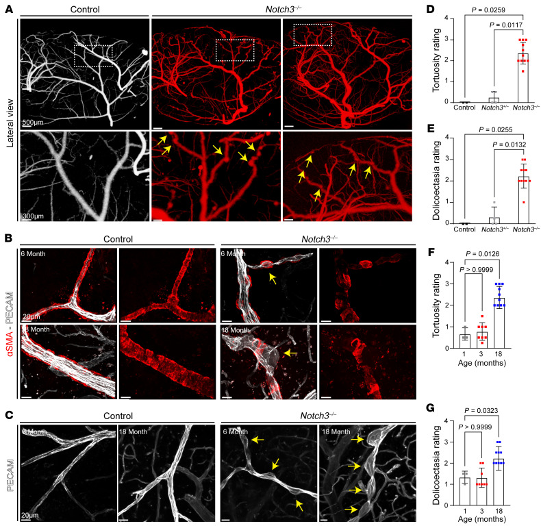
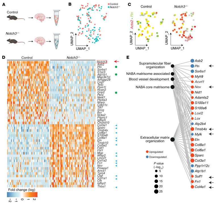
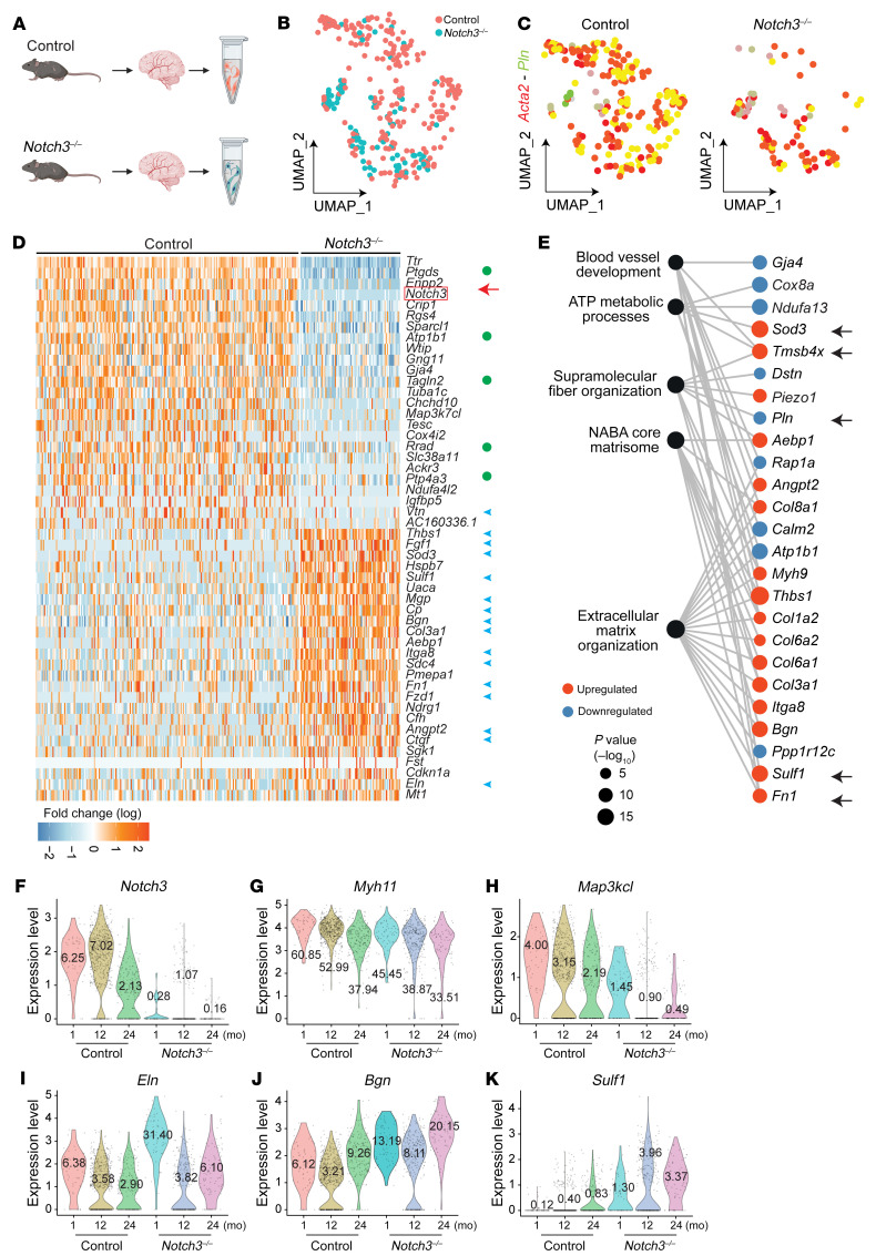
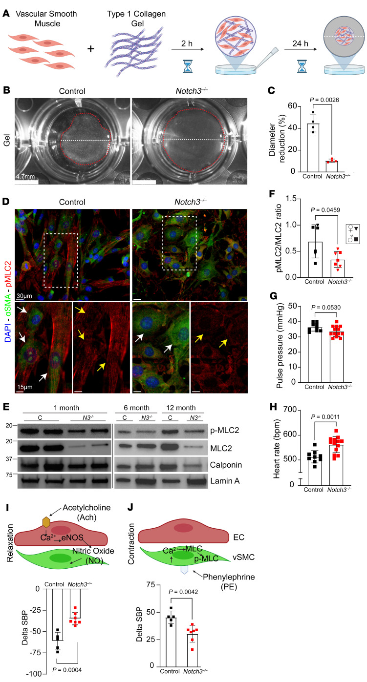
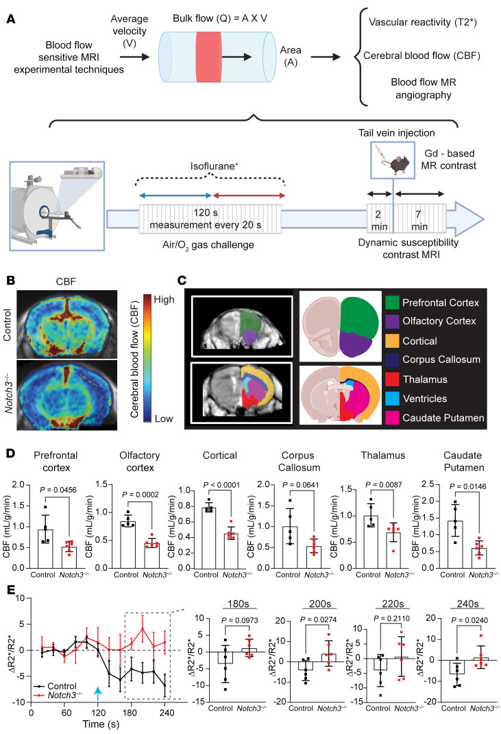
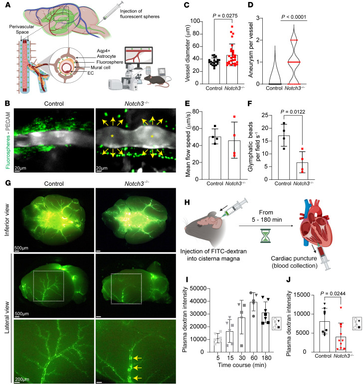
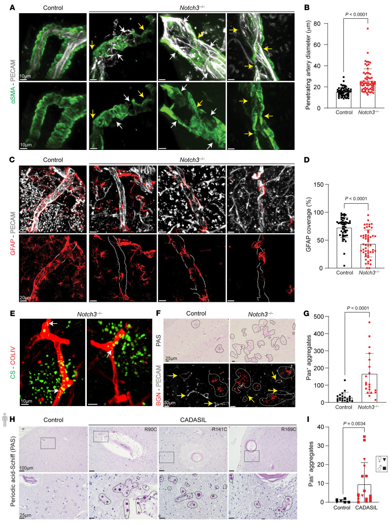
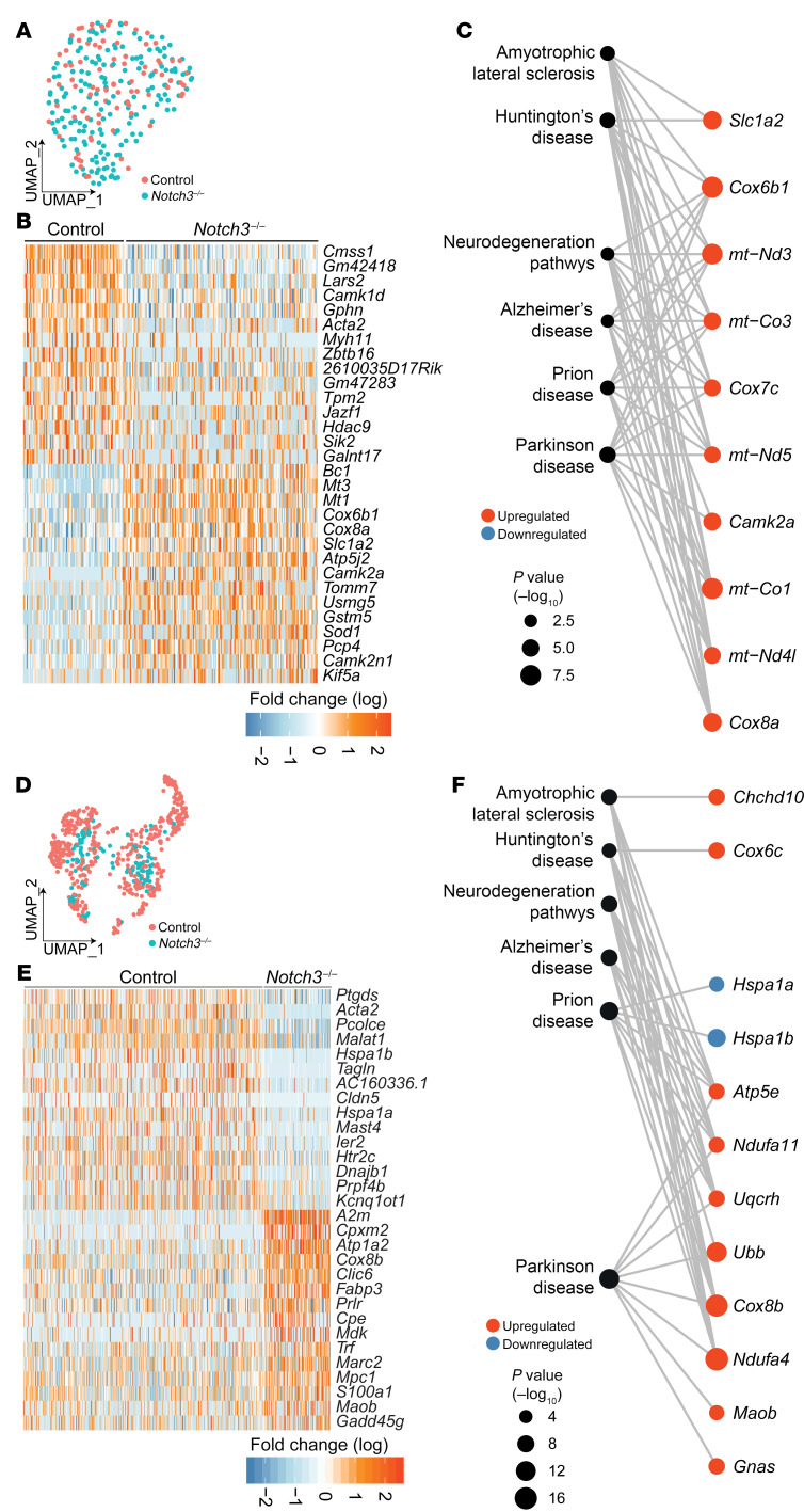
Vascular aging affects multiple organ systems, including the brain, where it can lead to vascular dementia. However, a concrete understanding of how aging specifically affects the brain vasculature, along with molecular readouts, remains vastly incomplete. Here, we demonstrate that aging is associated with a marked decline in Notch3 signaling in both murine and human brain vessels. To clarify the consequences of Notch3 loss in the brain vasculature, we used single-cell transcriptomics and found that Notch3 inactivation alters regulation of calcium and contractile function and promotes a notable increase in extracellular matrix. These alterations adversely impact vascular reactivity, manifesting as dilation, tortuosity, microaneurysms, and decreased cerebral blood flow, as observed by MRI. Combined, these vascular impairments hinder glymphatic flow and result in buildup of glycosaminoglycans within the brain parenchyma. Remarkably, this phenomenon mirrors a key pathological feature found in brains of patients with CADASIL, a hereditary vascular dementia associated with NOTCH3 missense mutations. Additionally, single-cell RNA sequencing of the neuronal compartment in aging Notch3-null mice unveiled patterns reminiscent of those observed in neurodegenerative diseases. These findings offer direct evidence that age-related NOTCH3 deficiencies trigger a progressive decline in vascular function, subsequently affecting glymphatic flow and culminating in neurodegeneration.

Keywords: Cardiovascular disease; Dementia; Neurological disorders; Neuroscience; Vascular Biology.


Conflict of interest statement

Conflict of interest: The authors have declared that no conflict of interest exists.

Figures

Figure 1
Figure 1. The aging vasculature experiences progressive loss of Notch3 that leads to ongoing disorganization, dedifferentiation, and detachment of VSMCs.
(A) Retinal vasculature from C57BL/6J mice at indicated ages. αSMA (green) identifies VSMCs, and PECAM (red) identifies endothelium. White arrows highlight VSMC loss. Scale bars: 200 μm (top row), 50 μm (bottom row). (B) Quantification of VSMC coverage at each time point from mixed-sex cohorts. Data are shown as the mean ± SD; n = 3–6. Welch’s t test. (C) Experimental design: Meningeal tissue and penetrating arteries were dissected from young (1 month) and aged (24 months) mice for scRNA-Seq. (D) Heatmap visualizing the top 50 differentially expressed genes (DEGs) in VSMCs. Green circles indicate genes that regulate muscle cell contraction. (EL) Violin plots from selected transcripts. (M) Human brain vessel sections stained with αSMA (green) to visualize smooth muscle and NOTCH3 (white). White arrows indicate NOTCH3 in VSMC nuclei. Scale bars: 2 μm. (N) Quantification of NOTCH3+ nuclei in human brain VSMCs (20–50 μm vessel diameter) at indicated age ranges. (O) NOTCH3 mean intensity per nuclei in human brain VSMCs. For N and O, data are shown as the mean ± SD; n = 43–53 vessels, 6–7 patients per age group. Kruskal-Wallis with multiple testing correction.
Figure 2
Figure 2. Loss of Notch3 in VSMCs and pericytes results in a decrease in transcripts that regulate contractility and an increase in transcripts associated with extracellular matrix.
(A) Schema of experimental design. Superficial brain vessels and penetrating brain arteries were dissected from mature (12-month-old) female and male Notch3–/– and littermate control mice and enzymatically dissociated to obtain single-cell suspensions for scRNA-Seq. n = 8 total mice, 2 female and 2 male pooled per genotype. (B) Uniform manifold approximation and projection (UMAP) plot of scRNA-Seq visualizing spread of data from VSMCs of the 2 genotypes. (C) Feature plot identifies Acta2+Pln+ VSMCs in control and Notch3–/–. (D) Heatmap visualizing the top 50 DEGs identified in VSMCs. (E) Gene Ontology enrichment of Notch3–/– VSMCs from the top 10 unique ontology categories and selected member genes within each category. Dot color indicates direction of expression change upon loss of Notch3–/–, while size indicates significance of enrichment. (F) UMAP plot of scRNA-Seq visualizing spread of data from pericytes of the 2 genotypes at 12 months of age. (G) Feature plot identifies Pdgfrb+Ogn+ pericytes in control and Notch3–/–. (H) Heatmap visualizing the top 50 DEGs identified in pericytes. (I) Gene Ontology enrichment of Notch3–/– pericytes from the top 10 unique ontology categories and selected member genes within each category. Dot color indicates direction of expression change upon loss of Notch3–/–, while size indicates significance of enrichment. Yellow stars indicate genes identified as DEGs in the same direction in both Notch3–/– VSMCs and Notch3–/– pericytes. For D and H, green circles indicate genes that regulate muscle cell contraction; blue arrowheads indicate ECM transcripts. Yellow stars indicate genes identified as DEGs in the same direction in both Notch3–/– VSMCs and Notch3–/– pericytes.
Figure 3
Figure 3. Notch3 deficiency leads to vascular dilation and tortuosity in the middle cerebral artery.
(A) Schema of experimental analysis whereby micro-CT overlay of the brain vasculature of Notch3–/– (red) and control (white) mice identifies abnormalities. (B) Coronal and inferior micro-CT overlay of Notch3–/– and control animals at 18 months. Yellow arrows indicate tortuosity and aneurysms in Notch3–/– compared with control. Scale bars: 1000 μm (left), 500 μm (right). (C) Schema of the regions of middle cerebral artery (MCA) measured in D. (D) Volumetric analysis of the MCA from the circle of Willis to 3,000 μm in Z axis (from the MCA branch point upward) in control and Notch3–/–. Maximum-intensity projections of MCA were color-coded by volume and overlaid on the base micro-CT visualization (white) in the lateral view. Yellow arrows indicate tortuosity. Scale bars: 500 μm. (E) Quantification of MCA volumes from Notch3–/– and control littermates at 18 months. n = 4–6; Mann-Whitney test. (F) Quantification of tortuosity index for the MCA in a cohort of control and Notch3–/– animals. n = 4–6; Welch’s t test.
Figure 4
Figure 4. Microaneurysms and tortuosity in Notch3–/– mice are associated with progressive loss and disorganization of VSMCs.
(A) Representative micro-CT images of microaneurysms in higher-order branches of the MCA. Yellow arrows indicate points of dilation. (B) Images illustrating microaneurysms and tortuosity in aging Notch3–/– brain penetrating arteries in the context of VSMC loss at 6 and 18 months as indicated. Yellow arrows indicate aneurysms in Notch3–/– animals. (C) Images of microaneurysms in aging Notch3–/– brain vessels. Yellow arrows point to dilations (PECAM, white). (D) Tortuosity rating in WT, heterozygous, and null mice. n = 3–11; Kruskal-Wallis test. (E) Dolichoectasia rating across three Notch3 genotypes. n = 3–11; Kruskal-Wallis test. (F) Tortuosity rating at 3 progressive time points in Notch3–/– animals. n = 4–10; Kruskal-Wallis test. (G) Dolichoectasia at 3 progressive time points in Notch3–/– animals. n = 4–10; Kruskal-Wallis test. For D and F, numbers indicate severity (1, zero to minimal vasculature tortuosity; 3, severe tortuosity across 7–15 vessels per animal). For E and G, numbers indicate severity (1, zero to minimal dolichoectasia; 3, severe dolichoectasia across 10–15 vessels per animal).
Figure 5
Figure 5. Transcriptional signature associated with Notch3 loss at 1 month in VSMCs.
(A) Graphical illustration of experimental design. Superficial brain vessels and penetrating brain arteries were dissected from 1-month Notch3–/– and littermate control mice and enzymatically dissociated to obtain single-cell suspensions for scRNA-Seq. (B) UMAP plot of scRNA-Seq visualizing spread of data from VSMCs of the 2 genotypes. (C) Feature plot identifies Acta2+Pln+ VSMCs in control and Notch3–/–. (D) Heatmap visualizing the top 50 DEGs identified in VSMCs. Green circles indicate genes that regulate muscle cell contraction; blue arrowheads indicate ECM transcripts. (E) Gene Ontology enrichment of Notch3–/– VSMCs for the top 5 unique ontology categories and selected member genes within each category. Dot color indicates the direction of expression change upon loss of Notch3 expression, while size indicates significance of enrichment. Arrows highlight gene products that were selected for validation using immunofluorescence (see Supplemental Figures).
Figure 6
Figure 6. Transcriptional signature associated with Notch3 deficiency at 24 months in VSMCs.
(A) Graphical illustration of experimental design. Superficial brain vessels and penetrating brain arteries were dissected from 24-month Notch3–/– and littermate control mice and enzymatically dissociated to obtain single-cell suspensions for scRNA-Seq. (B) UMAP plot of scRNA-Seq visualizing spread of data from VSMCs of the 2 genotypes. (C) Feature plot identifies Acta2+Pln+ VSMCs in control and Notch3–/–. (D) Heatmap visualizing the top 50 DEGs identified in VSMCs. Green circles indicate genes that regulate muscle cell contraction; blue arrowheads indicate ECM transcripts. (E) Gene Ontology enrichment of Notch3–/– VSMCs from the top 5 unique ontology categories and selected member genes within each category. Dot color indicates the direction of expression change upon loss of Notch3 expression, while size indicates significance of enrichment. Arrows highlight the gene products that were selected for validation by immunofluorescence (see Supplemental Figures). (FK) Violin plots from selected transcripts across ages and genotypes.
Figure 7
Figure 7. Notch3 deficiency results in vascular dysfunction due to impaired contractility.
(A) Diagram of experimental design. VSMCs from 1-month control and Notch3–/– mice were isolated and mixed with type I collagen to form a cellular hydrogel, detached, and observed 24 hours later. (B) Images of polymer gels from control and Notch3–/– cells after 24 hours. Note the differences in the contraction of the gel with regard to diameter (white dashed line) and total size of gel (red dashed line). (C) Quantification of percent gel diameter reduction 24 hours after plating. Bars indicate mean ± SD; n = 4 biological replicates; Welch’s t test. (D) Immunofluorescence of phosphorylated myosin light chain 2 (p-MLC2; red). White arrows highlight αSMA (green); yellow arrows highlight p-MLC2 (red). (E) VSMC-enriched lysates from mixed-sex cohorts of Notch3–/– and control aortae at the indicated ages were evaluated for expression of p-MLC2, MLC2, calponin, and lamin A (loading control). (F) Quantification of p-MLC2/MLC2 from a 1-month mixed-sex cohort. Data are shown as the mean ± SD; n = 6–7; Welch’s t test. Female data are indicated by inverted triangles, male data by squares. (G) Pulse pressure in 18-month-old male control and Notch3–/– mice. (H) Heart rate measured in 18-month male control and Notch3–/– mice. (I) Simplified diagram of the molecular pathway of acetylcholine-driven relaxation in VSMCs and quantification of systolic blood pressure response to acetylcholine in 18-month male control and Notch3–/– animals. (J) Simplified diagram of the molecular pathway of phenylephrine-driven contraction in VSMCs and quantification of systolic blood pressure response to phenylephrine in 18-month male control and Notch3–/– animals. For G and H, data are shown as the mean ± SD; n = 9–13, unpaired Student’s t test. For I and J, data are shown as the mean ± SD; n = 5–7, unpaired Student’s t test.
Figure 8
Figure 8. Notch3 deficiency delays cerebral vascular blood flow.
(A) Schematic depiction of the MRI techniques used to obtain blood flow parameters and timing of acquisition. (B) Representative MRI image with superimposed cerebral blood flow (CBF) parameters obtained from dynamic susceptibility contrast MRI for each group. (C) Regional map of the brain regions used for CBF measurements. (D) Quantification of CBF measurements at the indicated regions. (E) Average R2* MRI during oxygen challenge across control and Notch3–/– animals. The blue arrow indicates the time at which the switch between room air and oxygen occurred during the experimental design indicated in A. For D and E, n = 6 animals per group in each genotype; Welch’s t test. Data are shown as the mean ± SD (D) and ± SEM (E).
Figure 9
Figure 9. Notch3 deficiency delays glymphatic flow.
(A) Experimental design. Mice were anesthetized and injected with PECAM antibodies. A cannula inserted in the cisterna magna delivered fluorescent beads, which reached the perivascular space and were visualized by live imaging from the intact skull. (B) In vivo images of PECAM-labeled vessels in Notch3–/– and control. Arrows indicate flow of FITC-beads; asterisks indicate aneurysms. (C) Quantification of vessel diameter (from live images). Data are shown as the mean ± SD; n = 20 vessels, 5 animals (control), and 28 vessels, 8 animals (Notch3–/–), from mixed sexes at 6 months. Mann-Whitney test. (D) Quantification of aneurysms per vessel. n = 20 vessels, 5 animals (control), and 28 vessels, 8 animals (Notch3–/–), from mixed sexes at 6 months. One-sample Wilcoxon’s test. (E) Quantification of mean bead velocity. (F) Quantification of beads per visual field in mixed sexes at 6 months. Data are shown as the mean ± SD; n = 4; Welch’s t test for E and F. (G) Representative inferior and lateral images of Notch3–/– and control brains harvested 3 hours after cisternal injection. Yellow arrows indicate bead stagnation. (H) Experimental design. Mice were injected with FITC-dextran into the cisterna magna; blood was collected by cardiac puncture to assess fluorescence as a measurement of glymphatic clearance. (I) Quantification of fluorescence intensity after cisternal FITC-dextran injection across multiple time points in a mixed-sex cohort of 3-month C57BL/6J mice. n = 5–7 animals per time point and/or condition, as indicated in graph; unpaired Student’s t test. (J) Quantification of plasma fluorescence intensity 5 minutes after cisternal FITC-dextran injection in a mixed-sex cohort of 6-month Notch3–/– and control animals. Data are shown as the mean ± SD; n = 8–10 animals per time point and/or condition, as indicated; Welch’s t test. For I and J: squares, male; inverted triangles, female.
Figure 10
Figure 10. Deletion of Notch3 results in enlargement of penetrating arteries, detachment of astrocytes, and accumulation of extracellular proteoglycans in the brain parenchyma.
(A) Penetrating arteries of 24-month control and Notch3–/– mice stained with αSMA (green) and PECAM (white). White arrows indicate loss of VSMC coverage; yellow arrows indicate dilations and tortuosity. (B) Diameter of penetrating arteries. Data are shown as the mean ± SD; n = 10–16 vessels per animal, 5 mice per group. Mann-Whitney test. (C) Penetrating arteries of 24-month control and Notch3–/– mice stained with GFAP (red) and PECAM (white). White dotted lines highlight regions of poor GFAP+ coverage. (D) Percentage of vessel covered by GFAP+ astrocytes in control and Notch3–/–. Data are shown as the mean ± SD; n = 9–13 vessels per animal, 5 mice per group. Mann-Whitney test. (E) Vascular capillaries stained with type IV collagen (red) and chondroitin sulfate (green) in Notch3–/–. White arrows identify intracellular chondroitin sulfate. (F) Periodic acid–Schiff (PAS) staining in coronal sections of aged (24-month) control and Notch3–/–. Identification of biglycan (red). Yellow arrows indicate vessels; dashed white lines highlight biglycan. (G) Quantification of PAS+ aggregates in control and Notch3–/–. Data are shown as the mean ± SD; n = 5 visual fields per animal, 4 mice per genotype. Mann-Whitney test. (H) PAS staining in brain sections from control and CADASIL patients. Dotted black lines highlight PAS granules. (I) Average number of PAS+ aggregates in age-matched, mixed-sex cohort of control and CADASIL patient brain samples. Data are shown as the mean ± SD; n = 6–10 visual fields per individual, n = 6 control and 17 CADASIL patients. Mann-Whitney test. Squares, male; inverted triangles, female.
Figure 11
Figure 11. Deletion of Notch3 leads to progressive transcriptional alterations in the neuronal compartment, revealing neurodegeneration.
(A) UMAP plot of scRNA-Seq data from 12-month cortical neurons. (B) Heatmap of the top 30 DEGs in control and Notch3–/– cortical neurons. (C) Network of selected neurodegenerative disease–associated KEGG pathways of cortical neuron DEGs in Notch3–/–. (D) UMAP data from 24-month cortical neurons. (E) Heatmap of the top 30 DEGs in control and Notch3–/– cortical neurons at 24 months. (F) Network visualization of selected neurodegenerative disease–associated KEGG pathways and member genes from the top 20 enriched pathways.

References

    1. Sutton NR, et al. Molecular mechanisms of vascular health: insights from vascular aging and calcification. Arterioscler Thromb Vasc Biol. 2023;43(1):15–29. doi: 10.1161/ATVBAHA.122.317332. - DOI - PMC - PubMed
    1. Abdelkarim D, et al. A neural-vascular complex of age-related changes in the human brain: anatomy, physiology, and implications for neurocognitive aging. Neurosci Biobehav Rev. 2019;107:927–944. doi: 10.1016/j.neubiorev.2019.09.005. - DOI - PubMed
    1. Rajeev V, et al. Chronic cerebral hypoperfusion: a critical feature in unravelling the etiology of vascular cognitive impairment. Acta Neuropathol Commun. 2023;11(1):93. doi: 10.1186/s40478-023-01590-1. - DOI - PMC - PubMed
    1. Hattori Y, et al. Gradual carotid artery stenosis in mice closely replicates hypoperfusive vascular dementia in humans. J Am Heart Assoc. 2016;5(2):e002757. doi: 10.1161/JAHA.115.002757. - DOI - PMC - PubMed
    1. Claassen J, et al. Regulation of cerebral blood flow in humans: physiology and clinical implications of autoregulation. Physiol Rev. 2021;101(4):1487–1559. doi: 10.1152/physrev.00022.2020. - DOI - PMC - PubMed

LinkOut - more resources