. 2023 Dec 5;120(49):e2306390120.
doi: 10.1073/pnas.2306390120. Epub 2023 Nov 28.

Nucleolin binds to and regulates transcription of hepatitis B virus covalently closed circular DNA minichromosome

Affiliations

Nucleolin binds to and regulates transcription of hepatitis B virus covalently closed circular DNA minichromosome

Yuchen Xia et al. Proc Natl Acad Sci U S A. .

Abstract

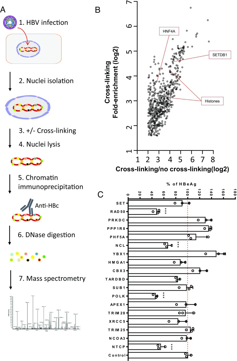
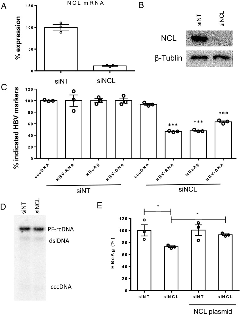
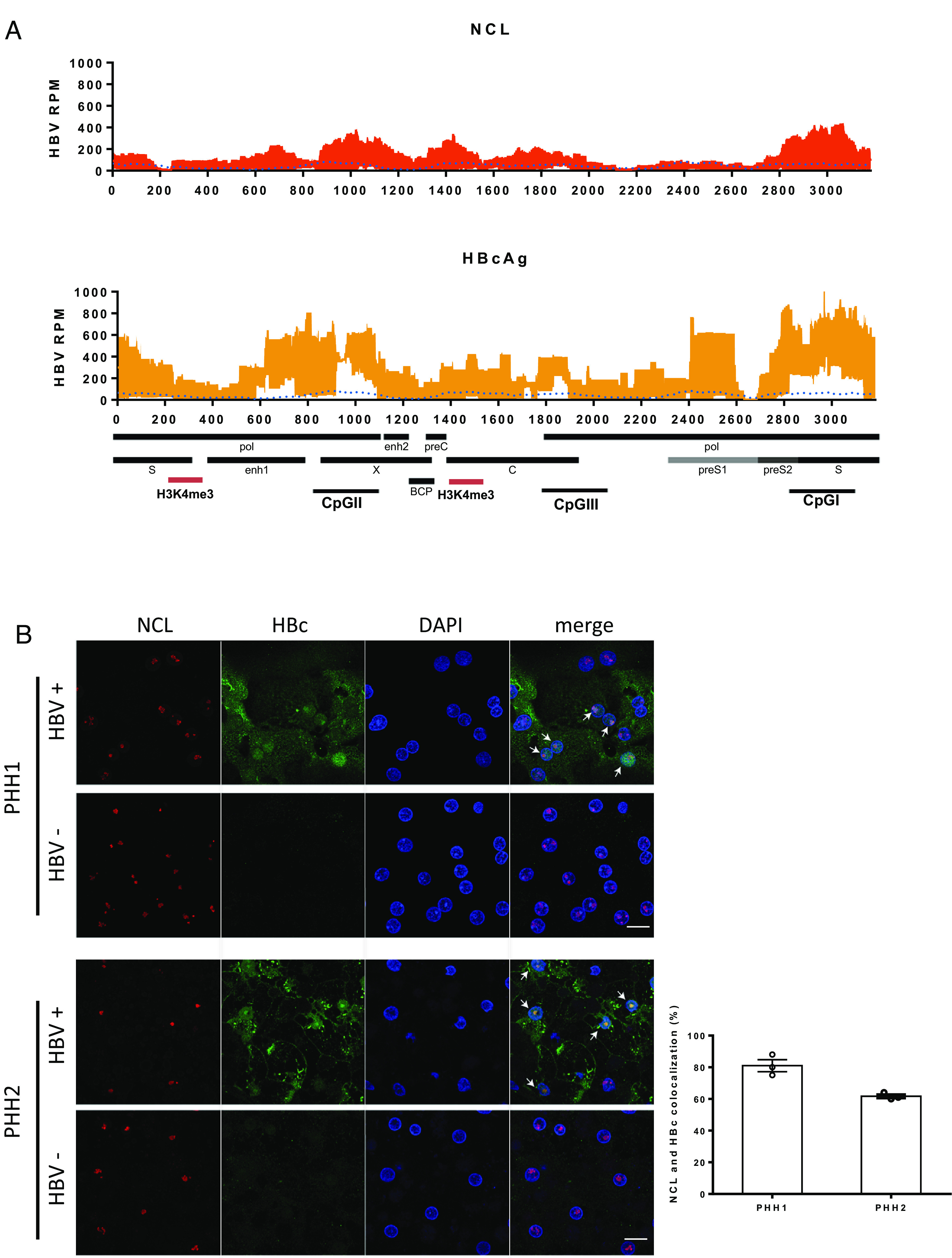
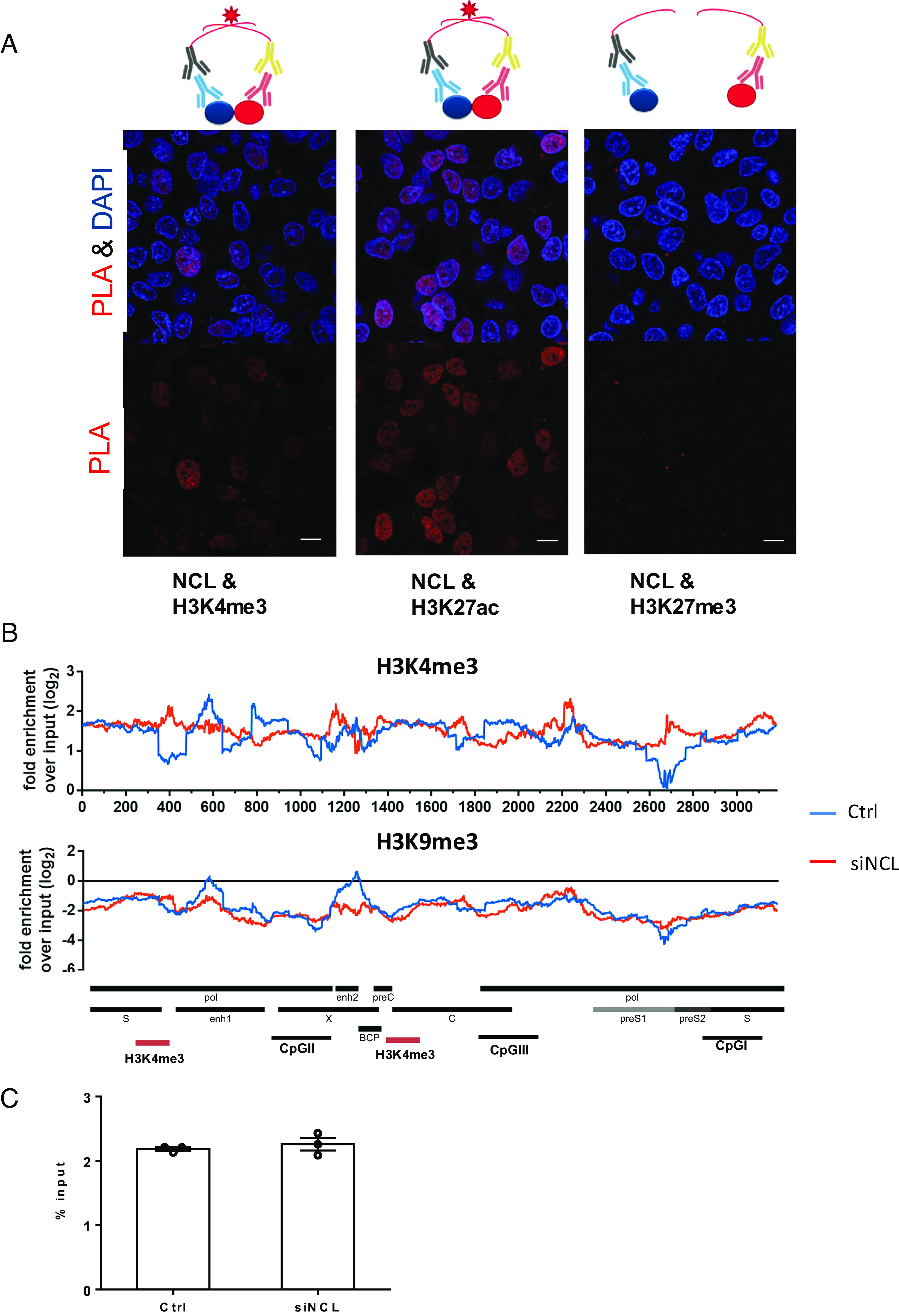
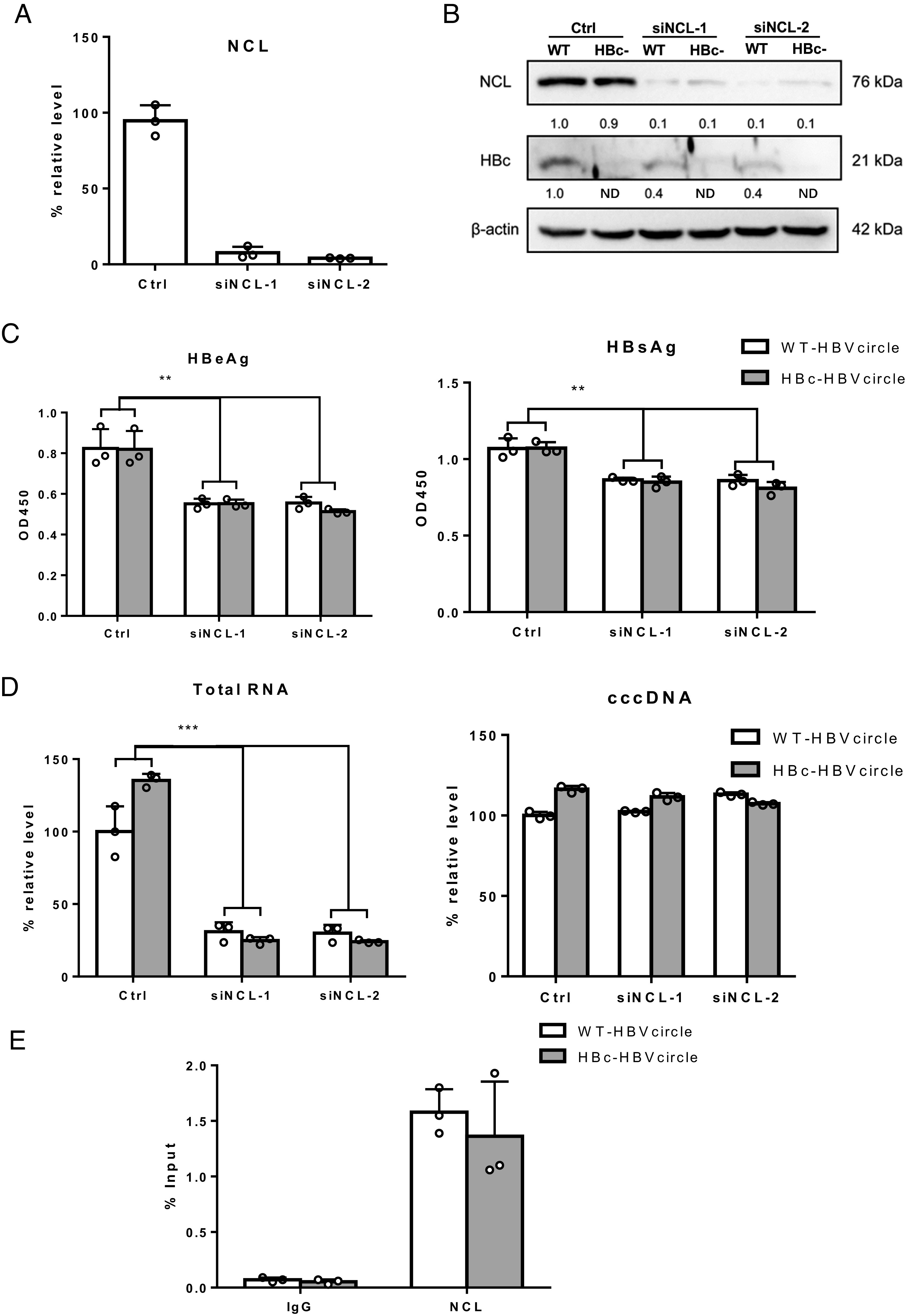
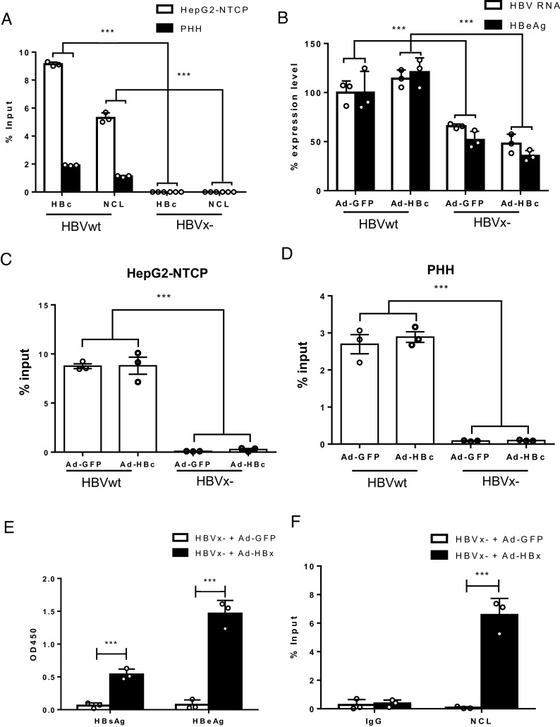
Hepatitis B virus (HBV) remains a major public health threat with nearly 300 million people chronically infected worldwide who are at a high risk of developing hepatocellular carcinoma. Current therapies are effective in suppressing HBV replication but rarely lead to cure. Current therapies do not affect the HBV covalently closed circular DNA (cccDNA), which serves as the template for viral transcription and replication and is highly stable in infected cells to ensure viral persistence. In this study, we aim to identify and elucidate the functional role of cccDNA-associated host factors using affinity purification and protein mass spectrometry in HBV-infected cells. Nucleolin was identified as a key cccDNA-binding protein and shown to play an important role in HBV cccDNA transcription, likely via epigenetic regulation. Targeting nucleolin to silence cccDNA transcription in infected hepatocytes may be a promising therapeutic strategy for a functional cure of HBV.

Keywords: HBV minichromosome; antiviral; epigenetics; proteomics; viral replication.


Conflict of interest statement

Competing interests statement:The authors declare no competing interest.

Figures

Fig. 1.
Fig. 1.
Identification of HBV cccDNA-associated proteins. (A) The workflow of HBV cccDNA-associated proteins isolation and identification. HepG2-NTCP cells were infected with HBV for 7 d and nuclei were isolated, either cross-linked and lysed or just lysed. Anti-HBc or unrelated antibody was used to specifically immunoprecipitate the cccDNA minichromosome. The resulting pulldown samples were digested with DNAse and proteins identified through mass spectrometry. Uninfected HepG2-NTCP cells were processed similarly as controls. (B) Result of mass spectrometry analysis. Fold enrichments over IgG control sample with or without cross-linking were plotted. In uninfected HepG2-NTCP cells, no specifically enriched cellular proteins were noted. (C) siRNA studies of 17 selected candidate genes in HBV-infected cells. siRNAs targeting the 17 genes and NTCP (positive control), nontargeting (siNT) control were transfected into HepG2-NTCP cells first and then infected with HBV 2 d later. Five days after infection, HBeAg levels were determined by ELISA. All data are normalized to siNT control sample (set as 100) and shown as means + SEM. The results are representative of three independent experiments. ***P < 0.001.
Fig. 2.
Fig. 2.
Characterization of nucleolin in HBV replication. siRNA against NCL were transfected into HepG2-NTCP cells; (A) mRNA and (B) protein levels of NCL were evaluated by qPCR (normalized to the siNT control) and western blot, respectively. (C) Five days after HBV infection, various HBV infection markers including cccDNA, HBV RNA (total), HBeAg, and HBV DNA in cell culture supernatant were determined and levels normalized to the siNT control. (D) The amount of HBV cccDNA was further determined by Southern blot. (E) HepG2-NTCP cells were transfected with siRNA against NCL or control and then transfected with or without plasmid overexpressing NCL. After HBV infection, HBeAg in cell culture supernatant were determined by ELISA. *P < 0.05 and ***P < 0.001.
Fig. 3.
Fig. 3.
Distribution of nucleolin on cccDNA and in HBV-infected cells. (A) Distribution of nucleosomes and protein binding sites were determined by cccDNA ChIP sequencing across the HBV genome. HBV cccDNA ChIP sequencing experiment was performed by using NCL or HBc antibody. Read density for each track is represented on the y-axis as HBV-derived reads per million total reads (HBV RPM). Averages and SDs from three independent experiments are shown. The signal of input is blotted as the blue dotted line. HBV transcripts, enhancer elements, CpG islands, basal core promoter (BCP), and highly enriched H3K4me3 binding sites (red) on HBV genome are schematically displayed below the x-axis. The results are representative of three independent experiments. (B) Confocal images of immunofluorescence staining for NCL and HBc. PHHs infected with or without HBV from two different donors were fixed and stained with antibodies against NCL (red) and HBc (green). The percentage of cells with NCL and HBc colocalization in HBc-positive cells was determined by counting three random images from each group. The results are representative of three independent experiments (Scale bar, 20 μm).
Fig. 4.
Fig. 4.
Interaction of nucleolin with HBV cccDNA. (A) Proximity ligation assay (PLA) to analyze the interaction of NCL and histones. The assay is schematically shown on the top of the figure. Antibodies against NCL or different histones were used in proximity ligation assay to determine their interaction in HBV-infected cells. HBV-infected HepG2-NTCP cells were fixed and incubated with NCL or histone antibodies. Secondary antibodies coupled with PLA probes were added and ligated. PLA signals are detected by fluorescent microscopy as discrete spots (in red) and provide the intracellular localization of the protein interaction (Scale bar, 10 μm). (B) Distribution of nucleosomes and H3K4me3 binding sites along the HBV genome in HBV-infected cells with or without NCL knockdown by ChIP-Seq. Read density for each track is represented by height on the y axis scaled to a minimum of 100 reads. Data are shown as fold-enrichment over input for each genome position, and various HBV genomic landmarks are displayed as in Fig. 3A. (C) The overall H3K4me3 signals on the HBV cccDNA were accessed by ChIP-qPCR and shown as % input.
Fig. 5.
Fig. 5.
The role of HBc in NCL-mediated transcriptional regulatory effect. (A) Huh7 cells were transfected with siRNA and 2 d later were transfected with WT-HBVcircle or HBc-HBVcircle. Three days later, NCL mRNA were evaluated by qPCR. (B) The expression of HBc and NCL protein was detected by western blot. (C) The levels of HBeAg and HBsAg in supernatants were measured by ELISA. (D) The relative levels of HBV RNA and HBVcircle DNA were determined by qPCR. (E) Chromatin immunoprecipitation was performed using lysates of Huh7 cells transfected with WT-HBVcircle or HBc-HBVcircle. Immunoprecipitation of HBVcircle using antibody against NCL or IgG control was analyzed by qPCR and shown as % input. **P < 0.01 and ***P < 0.001.
Fig. 6.
Fig. 6.
The role of HBx in NCL-mediated transcriptional regulatory effect. (A) Chromatin immunoprecipitation was performed using lysates of HepG2-NTCP and PHHs infected with wide-type HBV (HBVwt) or X- HBV (HBVx-). Immunoprecipitation of cccDNA minichromosome using antibody against HBc or NCL was analyzed by qPCR. The results are representative of three independent experiments. (B and C) HepG2-NTCP and (D) PHHs were infected with wide-type HBV (HBVwt) or X- HBV (HBVx-) and transduced with adenoviral vectors expressing HBc or GFP control. HBV RNA and HBeAg from infected HepG2-NTCP were determined. Immunoprecipitation of cccDNA minichromosome using antibody against NCL was analyzed by qPCR. (E and F) HepG2-NTCP cells were infection with X- HBV (HBVx-) and transduced with adenoviral vectors expressing HBx or GFP control the next day. Culture supernatant and cells were harvested on day 8 post-HBV infection. (E) HBsAg and HBeAg were determined by ELISA. (F) Immunoprecipitation of cccDNA using antibody against NCL was analyzed by qPCR and shown as % input (subtracting % input of IgG control). *P < 0.05, **P < 0.01, and ***P < 0.001.

References

    1. Hutin Y., et al. , Access to treatment for hepatitis B virus infection-worldwide, 2016. Am. J. Transplant. 18, 2595–2598 (2018). - PMC - PubMed
    1. Liang T. J., et al. , Present and future therapies of hepatitis B: From discovery to cure. Hepatology 62, 1893–1908 (2015). - PMC - PubMed
    1. Xia Y., Liang T. J., Development of direct-acting antiviral and host-targeting agents for treatment of hepatitis B virus infection. Gastroenterology 156, 311–324 (2018), 10.1053/j.gastro.2018.07.057. - DOI - PMC - PubMed
    1. Xia Y., Guo H., Hepatitis B virus cccDNA: Formation, regulation and therapeutic potential. Antiviral Res. 180, 104824 (2020). - PMC - PubMed
    1. Schreiner S., Nassal M., A role for the host DNA damage response in hepatitis B virus cccDNA formation-and beyond? Viruses 9, 125 (2017). - PMC - PubMed