. 2023 Dec 18;6(12):5502-5514.
doi: 10.1021/acsabm.3c00695. Epub 2023 Nov 28.

Modern Photodynamic Glioblastoma Therapy Using Curcumin- or Parietin-Loaded Lipid Nanoparticles in a CAM Model Study

Affiliations

Modern Photodynamic Glioblastoma Therapy Using Curcumin- or Parietin-Loaded Lipid Nanoparticles in a CAM Model Study

Jan Schulze et al. ACS Appl Bio Mater. .

Abstract

Natural photosensitizers, such as curcumin or parietin, play a vital role in photodynamic therapy (PDT), causing a light-mediated reaction that kills cancer cells. PDT is a promising treatment option for glioblastoma, especially when combined with nanoscale drug delivery systems. The curcumin- or parietin-loaded lipid nanoparticles were prepared via dual asymmetric centrifugation and subsequently characterized through physicochemical analyses including dynamic light scattering, laser Doppler velocimetry, and atomic force microscopy. The combination of PDT and lipid nanoparticles has been evaluated in vitro regarding uptake, safety, and efficacy. The extensive and well-vascularized chorioallantois membrane (CAM) of fertilized hen's eggs offers an optimal platform for three-dimensional cell culture, which has been used in this study to evaluate the photodynamic efficacy of lipid nanoparticles against glioblastoma cells. In contrast to other animal models, the CAM model lacks a mature immune system in an early stage, facilitating the growth of xenografts without rejection. Treatment of xenografted U87 glioblastoma cells on CAM was performed to assess the effects on tumor viability, growth, and angiogenesis. The xenografts and the surrounding blood vessels were targeted through topical application, and the effects of photodynamic therapy have been confirmed microscopically and via positron emission tomography and X-ray computed tomography. Finally, the excised xenografts embedded in the CAM were analyzed histologically by hematoxylin and eosin and KI67 staining.

Keywords: 3D cell culture; CAM model; drug delivery system; dual asymmetric centrifugation; lipid nanoparticles; natural photosensitizer.


Conflict of interest statement

The authors declare no competing financial interest.

Figures

Scheme 1
Scheme 1. Photophysical and Photochemical Processes during Photodynamic Therapy Illustrated in Combination with Pathways of PDT-Mediated Tumor Destruction
Parts of the image were drawn by using pictures from Servier Medical Art. Servier Medical Art by Servier is licensed under a Creative Commons Attribution 3.0 Unported License.
Figure 1
Figure 1
AFM images reveal the morphology of unloaded LNP and PS-loaded LNP in the height and amplitude modes. Scale bars represent 200 nm. Size distribution by number was measured by DLS and is displayed as histograms and frequency curves.
Figure 2
Figure 2
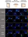
Transmission electron microscopy (TEM) micrographs of LNP, CUR LNP, and PTN LNP. Scale bars represent 500 nm in the upper row and 250 nm in the lower row.
Figure 3
Figure 3
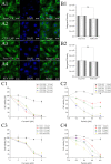
CLSM images of U87 glioblastoma cells after 4 h of incubation with PS-loaded LNP containing 30 μM curcumin or 1 μM parietin (A2, A4). Equivalent concentrations of free PS dissolved in medium containing 1% DMSO served as control (A1, A3). Scale bars represent 20 μm. Integrated density derived by CLSM data confirmed a homogeneous uptake for free and encapsulated curcumin (B1) and parietin (B2). Phototoxic effects of CUR LNP (C1), free CUR (C3), PTN LNP (C2), and free PTN (C4) were determined 24 h after irradiation (2.6, 5.3, 7.9, and 10.6 J/cm2) by MTT assays. Dark controls served as additional negative control (0 J/cm2).
Figure 4
Figure 4
Evaluation of the potential for irritation by HET-CAM. The top view image was captured on EDD 3 (after windowing eggs). On EDD 9, CAM was treated and observed continuously for 5 min, and any responses that occurred were recorded. The potential for irritation of unloaded LNP and PS-loaded LNP (without irradiation) was evaluated using the irritation score. NaCl 0.9% and NaOH 0.1 N served as control. Scale bars represent 1000 μm.
Figure 5
Figure 5
CAM images of successfully xenografted U87 glioblastoma cells were captured postapplication, preirradiation, and 1 h postirradiation. On EDD 12, xenografts were treated with CUR LNP and PTN LNP. Unloaded LNP and control (PBS) served as negative control. After 4 h of incubation, CAM was irradiated for 10 min by blue LED (13.2 J/cm2). Scale bars represent 1000 μm.
Figure 6
Figure 6
PET/CT images of the embryo with the treated xenografts. On EDD 13 (24 h postirradiation), 18F-FDG (8 ± 2 MBq per egg) was injected far away from xenografted tumors into a large blood vessel. White arrows indicate the locations of irradiated xenografts. Scale bars represent 2 mm.
Figure 7
Figure 7
HE and KI67 images of xenografts embedded in CAM and excised on EDD 14 (48 h postirradiation). Black arrows on KI67 images reveal spots of a high KI67 level, indicating high cell proliferation. Scale bars represent 200 μm.

References

    1. Zhao B.; Ma W. Extended adjuvant Temozolomide in newly diagnosed glioblastoma: the more, the better?. Neuro-oncology 2021, 23 (9), 1614–1615. 10.1093/neuonc/noab110. - DOI - PMC - PubMed
    1. Ostrom Q. T.; Cioffi G.; Waite K.; Kruchko C.; Barnholtz-Sloan J. S. CBTRUS Statistical Report: Primary Brain and Other Central Nervous System Tumors Diagnosed in the United States in 2014–2018. Neuro-oncology 2021, 23 (12 Suppl 3), iii1–iii105. 10.1093/neuonc/noab200. - DOI - PMC - PubMed
    1. Luo C.; Song K.; Wu S.; Hameed N. U. F.; Kudulaiti N.; Xu H.; Qin Z.-Y.; Wu J.-S. The prognosis of glioblastoma: a large, multifactorial study. British journal of neurosurgery 2021, 35 (5), 555–561. 10.1080/02688697.2021.1907306. - DOI - PubMed
    1. Rajaratnam V.; Islam M. M.; Yang M.; Slaby R.; Ramirez H. M.; Mirza S. P. Glioblastoma: Pathogenesis and Current Status of Chemotherapy and Other Novel Treatments. Cancers 2020, 12 (4), 937.10.3390/cancers12040937. - DOI - PMC - PubMed
    1. Buckner J. C.; Shaw E. G.; Pugh S. L.; Chakravarti A.; Gilbert M. R.; Barger G. R.; Coons S.; Ricci P.; Bullard D.; Brown P. D.; Stelzer K.; Brachman D.; Suh J. H.; Schultz C. J.; Bahary J.-P.; Fisher B. J.; Kim H.; Murtha A. D.; Bell E. H.; Won M.; Mehta M. P.; Curran W. J. Radiation plus Procarbazine, CCNU, and Vincristine in Low-Grade Glioma. New England journal of medicine 2016, 374 (14), 1344–1355. 10.1056/NEJMoa1500925. - DOI - PMC - PubMed

Publication types

LinkOut - more resources