. 2023 Nov 28;11(11):e007802.
doi: 10.1136/jitc-2023-007802.

MDSCs-derived GPR84 induces CD8+ T-cell senescence via p53 activation to suppress the antitumor response

Affiliations

MDSCs-derived GPR84 induces CD8+ T-cell senescence via p53 activation to suppress the antitumor response

Jinyan Liu et al. J Immunother Cancer. .

Abstract

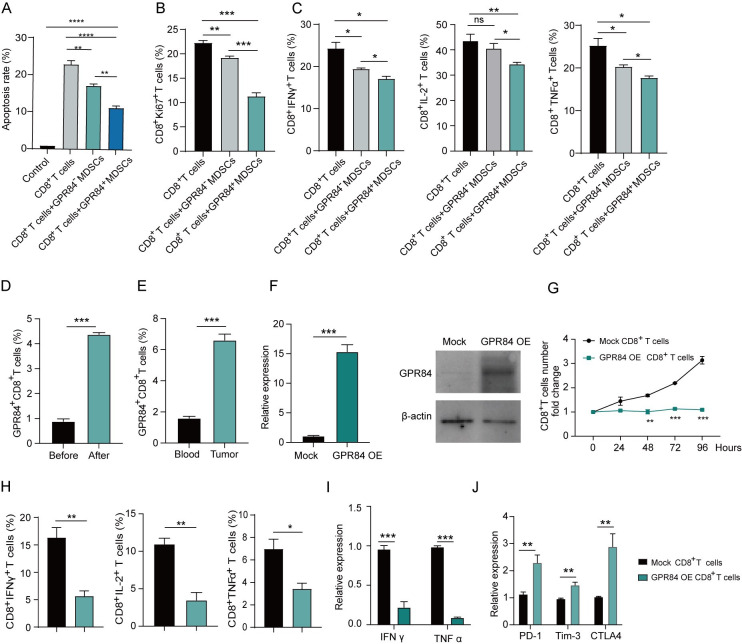
Backgrounds: G-protein-coupled receptor 84 (GPR84) marks a subset of myeloid-derived suppressor cells (MDSCs) with stronger immunosuppression in the tumor microenvironment. Yet, how GPR84 endowed the stronger inhibition of MDSCs to CD8+ T cells function is not well established. In this study, we aimed to identify the underlying mechanism behind the immunosuppression of CD8+ T cells by GPR84+ MDSCs.

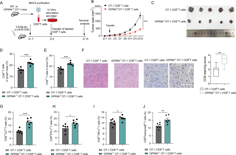
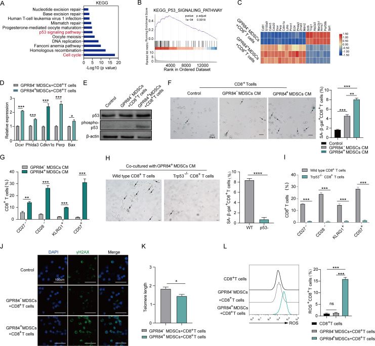
Methods: The role and underlying mechanism that MDSCs or exosomes (Exo) regulates the function of CD8+ T cells were investigated using immunofluorescence, fluorescence activating cell sorter (FACS), quantitative real-time PCR, western blot, ELISA, Confocal, RNA-sequencing (RNA-seq), etc. In vivo efficacy and mechanistic studies were conducted with wild type, GPR84 and p53 knockout C57/BL6 mice.

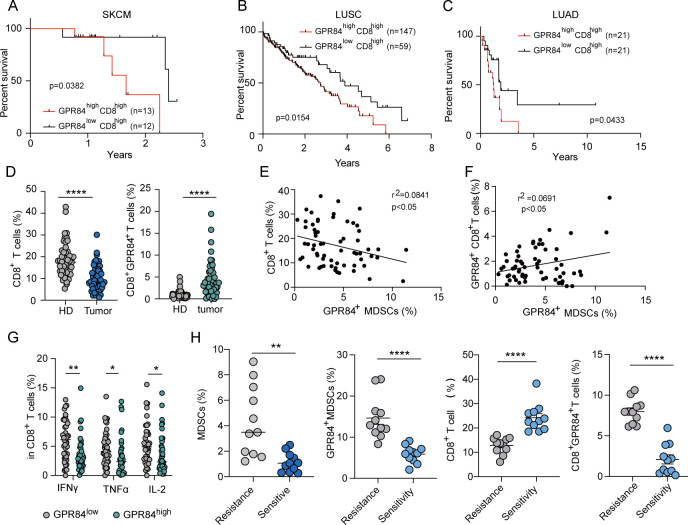
Results: Here, we showed that the transfer of GPR84 from MDSCs to CD8+ T cells via the Exo attenuated the antitumor response. This inhibitory effect was also observed in GPR84-overexpressed CD8+ T cells, whereas depleting GPR84 elevated CD8+ T cells proliferation and function in vitro and in vivo. RNA-seq analysis of CD8+ T cells demonstrated the activation of the p53 signaling pathway in CD8+ T cells treated with GPR84+ MDSCs culture medium. While knockout p53 did not induce senescence in CD8+ T cells treated with GPR84+ MDSCs. The per cent of GPR84+ CD8+ T cells work as a negative indicator for patients' prognosis and response to chemotherapy.

Conclusions: These data demonstrated that the transfer of GPR84 from MDSCs to CD8+ T cells induces T-cell senescence via the p53 signaling pathway, which could explain the strong immunosuppression of GPR84 endowed to MDSCs.

Keywords: CD8+T cells; GPR84; MDSCs; exosome; p53 signaling pathway.


Conflict of interest statement

Competing interests: No, there are no competing interests.

Figures

Figure 1
Figure 1
MDSCs inhibits CD8+ T-cell cytotoxicity depends on the GPR84 transfer. (A) The apoptotic rates of B16-OVA cells co-cultured with OT-I CD8+ T cells and GPR84+/– MDSCs for 6 hours (CD8+ T cells: MDSCs: tumor cells=1:4:1). CD8+ T cells and GPR84+/− MDSCs were co-cultured for 24 hours. Flow cytometry was used to analyze the proliferation (B) and function (C) of CD8+ T cells (CD8+ T cells: MDSCs=1:4). (D) The percentage of GPR84+ CD8+ T cells before and after being co-cultured with MDSCs (CD8+ T cells: MDSCs=1:4) was examined using flow cytometry. (E) The ratio of GPR84+ CD8+ T cells in blood and tumor tissues was detected in mice models bearing LLC cells. (F) CD8+ T cells were purified from C57 mice spleens after activation with an anti-CD3/CD28 antibody; the GPR84 plasmid was transfected, and GPR84-overexpressed CD8+ T cells were constructed. The transfection efficiency was examined using qRT-PCR and western blotting. Flow cytometry was used to analyze the proliferation (G) and function (H) of mock or GPR84-overexpressed CD8+ T cells. (I–J) The expression levels of functional and exhaustion-related genes on GPR84 overexpressed or mock CD8+ T cells were examined using qRT-PCR. Data were represented in at least three independent experiments. Data were presented as the mean±SEM. ns, not significant, *p<0.05, **p<0.01, ***p<0.001, ****p<0.0001. GPR84, G-protein-coupled receptor 84; MDSC, myeloid-derived suppressor cell; qRT-PCR, quantitative real-time PCR.
Figure 2
Figure 2
Loss of GPR84 enhances antitumor immunity of CD8+ T cells. GPR84, G-protein-coupled receptor 84; MDSC, myeloid-derived suppressor cell.
Figure 3
Figure 3
GPR84 suppresses the infiltration and reduces the antitumor ability of CD8+T cells. GPR84, G-protein-coupled receptor 84; IFN, interferon; IL, interleukin; MACS, magnet-activated cell sorting; s.c., subcutaneous under the skin; WT, wild type.
Figure 4
Figure 4
MDSCs transfer GPR84 to CD8+ T cells in an exosome-dependent way to repress CD8+ T-cell function. (A) After co-culturing with WT or GPR84-/-MDSCs for 48 hours, the expression of GPR84 on CD8+ T cells was determined using quantitative real-time PCR (qRT-PCR). (B–C) CD8+ T cells were purified from GPR84-KO mice and co-cultured with GPR84+ MDSCs. Then, flow cytometry (B) and imaging flow cytometry (C) were used to analyze the expression of GPR84 on CD8+ T cells. (D–E) Co-cultured CD8+ T cells with WT or GPR84–/– MDSCs directly (D) or indirectly (E). Flow cytometry was used to analyze the percentage of GPR84+CD8+ T cells at 48 hours and 72 hours. (F) CD8+ T cells were cultured in the cultured supernatant of WT or GPR84–/– MDSCs. Flow cytometry and imaging flow cytometry were used to analyze the expression of GPR84 on CD8+ T cells. (G) Flow cytometry was used to analyze the percentage of GPR84+ MDSCs after co-culturing with or without CD8+ T cells. (H) GPR84+ MDSCs were pretreated with GW4869 for 2 hours and then co-cultured with CD8+ T cells. After 48 hours, the percentage of GPR84+CD8+ T cells was analyzed using flow cytometry. (I) The morphology of exosomes obtained from WT or GPR84–/– MDSCs was detected using electron microscopy. The size distribution of the exosomes obtained from WT or GPR84–/– MDSCs was measured by using NTA assay. (J) The expression levels of Tsg101, CD9, GPR84, and GAPDH in MDSCs and MDSC-derived exosomes were examined using western blotting. (K) Representative images of PKH-67 (green) on CD8+ T cells co-cultured for 24 hours with PKH-67-tagged MDSC-derived exosomes. Nucleus was counter-stained with Hoechst (blue). (Bar, 10 µm). (L) CD8+ T cells were co-cultured with WT or GPR84–/– MDSCs-derived exosome for 24 hours. Flow cytometry was used to analyze the expression of GPR84 on CD8+ T cells. (M) CD8+ T cells were co-cultured with GPR84 antibody-tagged MDSCs for 24 hours. Then, flow cytometry was used to analyze the expression of GPR84 on CD8+ T cells. (N) GPR84+ MDSCs were pretreated with GW4869 for 2 hours and then co-cultured with CD8+ T cells. The killing ability (N) proliferation (O) and function-related cytokines (P) of CD8+ T cells were analyzed using flow cytometry. Data were represented in at least three independent experiments. Data were presented as the mean±SEM. ns, not significant, *p<0.05, **p<0.01, ***p<0.001, ****p<0.0001. GPR84, G-protein-coupled receptor 84; IFN, interferon; IL, interleukin; MDSC, myeloid-derived suppressor cell; NTA, nanoparticle tracking analysis; WT, wild type.
Figure 5
Figure 5
GPR84 induces the senescence of CD8+ T cells via the p53 pathway. CM, cultured medium; DAPI, 4,6-diamino-2-phenyl indole; GPR84, G-protein-coupled receptor 84; KEGG, Kyoto Encyclopedia of Genes and Genomes; MDSC, myeloid-derived suppressor cell; ROS, reactive oxygen species.
Figure 6
Figure 6
Clinical significance of GPR84 levels in CD8+ T cells. (A–C) Overall survival of skin cutaneous melanoma (SKCM), lung adenocarcinoma (LUAD), and lung squamous cell cancer (LUSC) was analyzed in GPR84high CD8high and GPR84low CD8high groups based on the data from The Cancer Genome Atlas. (D) The percentage of CD8+ T and GPR84+ CD8+ T cells in peripheral blood mononuclear cells from healthy donors (n=50) and patients with cancer (n=50) was analyzed using flow cytometry. (E) The correlation between CD8+ T cells and GPR84+ MDSCs was analyzed based on the flow cytometry data from healthy donors (n=63). (F) The correlation between GPR84+ CD8+ T cells and GPR84+ MDSCs was analyzed based on the flow cytometry data from healthy donors (n=63). (G) The expression of function-related markers in GPR84high and GPR84low CD8+ T cells was analyzed using flow cytometry from healthy donors (n=63). (H) The percentages of MDSCs, GPR84+ MDSCs, CD8+ T cells, and GPR84+ CD8+ T cells were analyzed in chemotherapy-resistant (n=11) or sensitive (n=11) patients with cancer using flow cytometry. Data were represented in at least three independent experiments. Data were presented as the mean±SEM. ns, not significant, *p<0.05, **p<0.01, ***p<0.001, ****p<0.0001. GPR84, G-protein-coupled receptor 84; HD, healthy donor; IFN, interferon; IL, interleukin; MDSC, myeloid-derived suppressor cell; TNF, tumor necrosis factor.

References

    1. Kim W, Chu TH, Nienhüser H, et al. . PD-1 signaling promotes tumor-infiltrating myeloid-derived Suppressor cells and gastric tumorigenesis in mice. Gastroenterology 2021;160:781–96. 10.1053/j.gastro.2020.10.036 - DOI - PMC - PubMed
    1. Myeloid-derived Itaconate suppresses antitumor immunity. Cancer Discov 2022:OF1. - PubMed
    1. Halaby MJ, Hezaveh K, Lamorte S, et al. . GCN2 drives macrophage and MDSC function and immunosuppression in the tumor microenvironment. Sci Immunol 2019;4:eaax8189. 10.1126/sciimmunol.aax8189 - DOI - PMC - PubMed
    1. Mohammadpour H, MacDonald CR, McCarthy PL, et al. . 2-adrenergic receptor signaling regulates metabolic pathways critical to myeloid-derived suppressor cell function within the TME. Cell Rep 2021;37:109883. 10.1016/j.celrep.2021.109883 - DOI - PMC - PubMed
    1. Ajith A, Mamouni K, Horuzsko DD, et al. . Targeting TREM1 augments antitumor T cell immunity by inhibiting myeloid-derived suppressor cells and restraining anti-PD-1 resistance. J Clin Invest 2023;133:e167951. 10.1172/JCI167951 - DOI - PMC - PubMed

Publication types

Substances