. 2023 Nov 27;13(1):20903.
doi: 10.1038/s41598-023-47394-0.

Cryopreservation of rat embryos at all developmental stages by small-volume vitrification procedure and rapid warming in cryotubes

Affiliations

Cryopreservation of rat embryos at all developmental stages by small-volume vitrification procedure and rapid warming in cryotubes

Shinsuke Seki et al. Sci Rep. .

Abstract

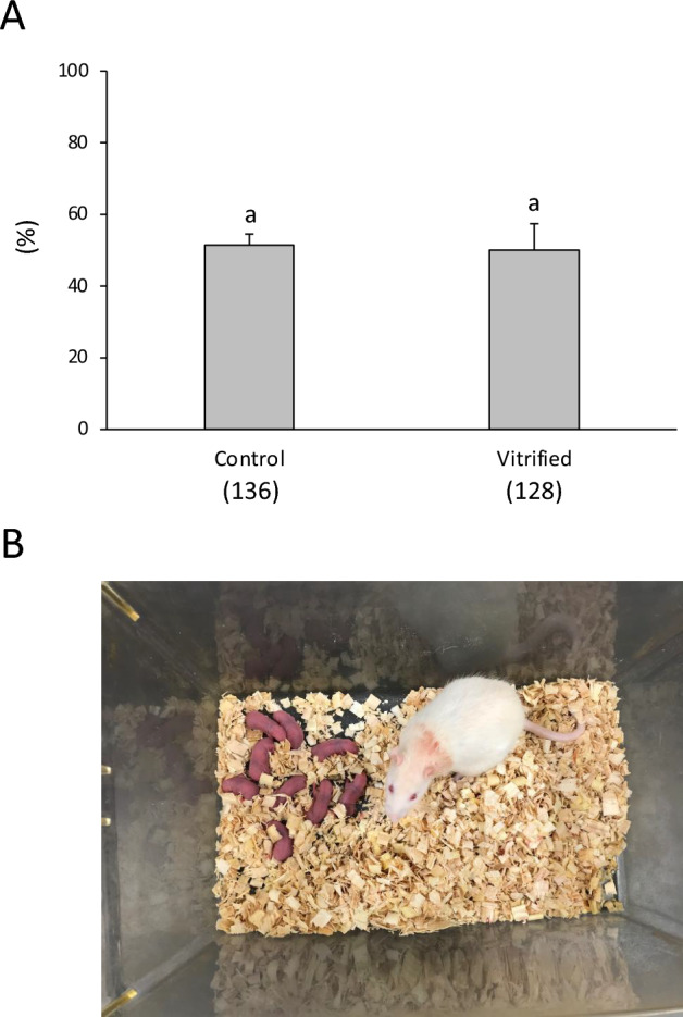
Intracellular ice formation during cryopreservation is lethal to the cell, including during warming. Here, we examined the effect of sample volume and warming rate on the cryopreservation success of 1-cell rat embryos based on successful development into blastocysts in vitro and to term in vivo following embryo transfer. Embryos were equilibrated in 5% propylene glycol solution for 10 min, held for 40 s at 0 °C in cryopreservation solution (5%PG + PEPeS), and cooled by immersion in liquid nitrogen. When 1-cell embryos were cryopreserved in a volume of 30-100 μL at a cooling rate of 5830-7160 °C/min and warmed at 35,480-49,400 °C/min by adding 1 mL of 0.3 M sucrose solution at 50 °C, 17.3-28.8% developed into blastocysts, compared with 57.0% of untreated embryos. However, when 1-cell embryos were cryopreserved in a smaller volume of 15 μl at 7950 °C/min and warmed at 68,850 °C/min, 58.8 ± 10.6% developed into blastocysts and 50.0 ± 7.4% developed to term, comparable to that of non-treated embryos (57.0 ± 5.4% and 51.4 ± 3.1%, respectively). Cryopreserved embryos at other developmental stages also showed high in vitro culture potential similar to that of the control. Using a conventional cryotube and a small-volume vitrification procedure with rapid warming, we achieved high levels of subsequent rat embryonic development at all developmental stages.


Conflict of interest statement

The authors declare no competing interests.

Figures

Figure 1
Figure 1
Survival and in vitro development of vitrified one-cell embryos (15 μL sample volume) warmed with sucrose solution at 23, 37, or 50 °C. The warming rates were 29,930 °C/min, 38,780 °C/min, and 68,850 °C/min, respectively. Each bar indicates the percentage of embryos surviving (open bars), developing to the two-cell stage (shaded bars), and developing to the blastocyst stage (closed bars) after cryopreservation (N = 8–10 experimental runs); numbers in parentheses indicate the number of one-cell embryos examined. Data are shown as means ± SEM. Different lowercase letters indicate a significant difference at P < 0.05.
Figure 2
Figure 2
Survival and in vitro development of cryopreserved one-cell embryos in a sample volume of 15, 30, 50, or 100 μL, which were cooled using direct immersion in liquid nitrogen (LN2) at a rate of 7950, 7160, 6800 and 5830 °C/min, respectively, and warmed with sucrose solution at 50 °C. The warming rates were 68,850, 49,400, 44,500 and 35,480 °C/min, respectively. Each bar indicates the percentage of embryos surviving (open bars), developing to the two-cell stage (shaded bars), and developing to the blastocyst (closed bars) after cryopreservation (N = 8–10 experimental runs); numbers in parentheses indicate the number of one-cell embryos examined. Data are shown as means ± SEM. Different lowercase letters indicate a significant difference at P < 0.05.
Figure 3
Figure 3
Survival and in vitro development of vitrified embryos at the 2-, 4-, and 8-cell embryos, morulae, and blastocysts in a sample volume of 15 μL, which were cooled using direct immersion in LN2 at a rate of 7950 °C/min, and warmed with sucrose solution at 50 °C at a rate of 68,850 °C/min. Each bar indicates the percentage of embryos surviving (open bars) and developing to the blastocyst stage (closed bars) after cryopreservation (N = 8–10 experimental runs); numbers in parentheses indicate the number of embryos examined at each developmental stage. Data are shown as means ± SEM. Different lowercase letters indicate a significant difference at P < 0.05. The developmental ability of blastocysts was assessed by determining whether they could develop into expanded blastocysts.
Figure 4
Figure 4
In vivo development of cryopreserved one-cell rat embryos. The one-cell embryos were vitrified with 5% PG for 10 min and PEPeS for 40 s. The embryos were warmed using 1 mL of sucrose solution at 50 °C. The cryopreserved or fresh embryos were transferred into the oviducts of pseudopregnant female rats. (A) Offspring survival. Numbers in parentheses indicate the number of one-cell embryos examined. Data are shown as means ± SEM. Different lowercase letters indicate a significant difference at P < 0.05. (B) Live pups derived from vitrified one-cell embryos.
Figure 5
Figure 5
(A) Thermal cycle oscilloscope trace of a 15 μL aqueous sample (PEPeS) in vitrification solution in a cryotube equipped at the bottom with a 50 μm Cu-constantan thermocouple. The cryotube sample was rapidly moved from room-temperature air (~ 23 °C) to LN2 and held for 5 s, and then held in air at 23 °C for 1 min before warming by the addition of 1 mL of sucrose solution at 50 °C. (B) Details of events during warming when the sample was warmed by adding 1 mL of sucrose solution at 50 °C. A straight line visually fit to the data revealed a warming rate of 84,000 °C/min. The temperature sampling rate was 500 readings/sec.
Figure 6
Figure 6
Cryopreservation and warming procedure for rat embryos developed in this study.

References

    1. Whittingham DG, et al. Survival of mouse embryos frozen to − 196 and − 269°C. Science. 1972;178:411–414. doi: 10.1126/science.178.4059.411. - DOI - PubMed
    1. Rall WF, Fahy GM. Ice-free cryopreservation of mouse embryos frozen to − 196°C by vitrification. Nature. 1985;313:573–575. doi: 10.1038/313573a0. - DOI - PubMed
    1. Seki S, Mazur P. Kinetics and activation energy of recrystallization of intracellular ice in mouse oocytes subjected to interrupted rapid cooling. Cryobiology. 2008;56:171–180. doi: 10.1016/j.cryobiol.2008.02.001. - DOI - PMC - PubMed
    1. Boutron P, Mehl P. Theoretical prediction of devitrification tendency: Determination of critical warming rates without using expansions. Cryobiology. 1990;27:359–377. doi: 10.1016/0011-2240(90)90015-V. - DOI - PubMed
    1. Fahy GM. Biological effects of vitrification and devitrification. In: Pegg DE, Karow AM, editors. The Biophysics of Organ Cryopreservation. Plenum Press; 1987. pp. 265–297.

Substances

LinkOut - more resources