Clinical Trial
. 2024 Jul;51(8):2384-2394.
doi: 10.1007/s00259-023-06525-5. Epub 2023 Nov 29.

MET-receptor targeted fluorescent imaging and spectroscopy to detect multifocal papillary thyroid cancer

Affiliations
Clinical Trial

MET-receptor targeted fluorescent imaging and spectroscopy to detect multifocal papillary thyroid cancer

Madelon J H Metman et al. Eur J Nucl Med Mol Imaging. 2024 Jul.

Abstract

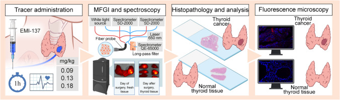
Purpose: Multifocal disease in PTC is associated with an increased recurrence rate. Multifocal disease (MD) is underdiagnosed with the current gold standard of pre-operative ultrasound staging. Here, we evaluate the use of EMI-137 targeted molecular fluorescence-guided imaging (MFGI) and spectroscopy as a tool for the intra-operative detection of uni- and multifocal papillary thyroid cancer (PTC) aiming to improve disease staging and treatment selection.

Methods: A phase-1 study (NCT03470259) with EMI-137 was conducted to evaluate the possibility of detecting PTC using MFGI and quantitative fiber-optic spectroscopy.

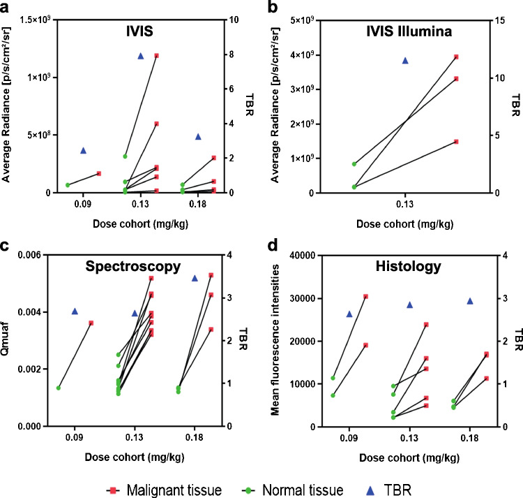
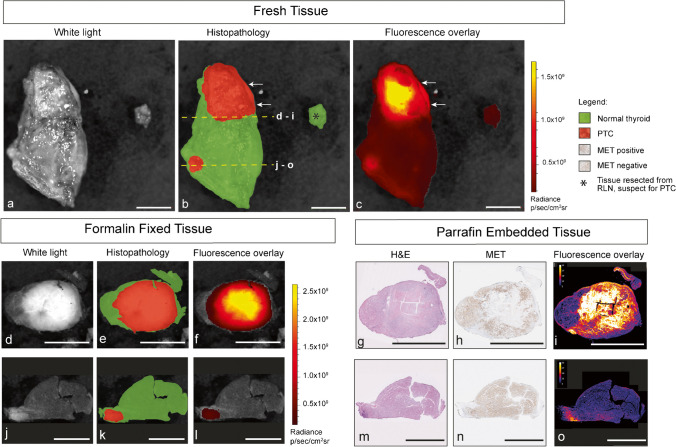
Results: Fourteen patients underwent hemi- or total thyroidectomy (TTX) after administration of 0.09 mg/kg (n = 1), 0.13 mg/kg (n = 8), or 0.18 mg/kg (n = 5) EMI-137. Both MFGI and spectroscopy could differentiate PTC from healthy thyroid tissue after administration of EMI-137, which binds selectively to MET in PTC. 0.13 mg/kg was the lowest dosage EMI-137 that allowed for differentiation between PTC and healthy thyroid tissue. The smallest PTC focus detected by MFGI was 1.4 mm. MFGI restaged 80% of patients from unifocal to multifocal PTC compared to ultrasound.

Conclusion: EMI-137-guided MFGI and spectroscopy can be used to detect multifocal PTC. This may improve disease staging and treatment selection between hemi- and total thyroidectomy by better differentiation between unifocal and multifocal disease.

Trial registration: NCT03470259.

Keywords: Molecular fluorescence–guided imaging; Multifocality; Papillary thyroid cancer; Spectroscopy.


Conflict of interest statement

GMvD is founder, shareholder, and CEO of TRACER Europe BV (Groningen, the Netherlands). None of the other authors reported any potential conflicts of interest.

Figures

Fig. 1
Fig. 1
Study workflow illustrating the steps for tracer administration, imaging, analysis, and validation of in vivo tracer binding following intravenous administration. Abbreviations: EMI-137, investigational medicinal product; h, hour; mg, milligram; MFGI, molecular fluorescence–guided imaging; kg, kilogram
Fig. 2
Fig. 2
a An overview of fluorescent intensities per dosage cohort of formalin-fixed papillary thyroid cancer (PTC) and healthy thyroid tissue imaged with the IVIS Spectrum, the IVIS Lumina (b), and spectroscopy (c). The mean fluorescence intensity per dosage cohort acquired from histological slides is provided in d. Green dots resemble healthy thyroid tissue, red squares resemble malignant tissue, and blue triangles resemble the TBR per dosage cohort (depicted on the right y-axis)
Fig. 3
Fig. 3
Representative images in the 0.13 mg/kg dosage cohort of a patient without detection of PTC multifocality on pre-operative ultrasound. The right thyroid lobe is imaged with the IVIS Spectrum as a fresh specimen from a dorsal perspective (a). The location of PTC foci was derived from the final histopathology results and drawn as a layer on the imaged fresh specimen (b). The primary tumor is in the cranial part of the right thyroid lobe. A second fluorescent focus located close to the ligament of Berry is visualized and confirmed as multifocal PTC on histopathological assessment (mo). The asterisk marks a piece of tissue clinically suspect for PTC and resected from the recurrent laryngeal nerve. The fluorescence intensity of this tissue was comparable to healthy thyroid tissue (b, c). Histopathological assessment of the resected tissue from the RLN showed healthy thyroid tissue. The region between the two arrows indicates a region with a higher fluorescence signal outside the tumor, suggesting a tumor deposition (b, c). The histopathological assessment showed a positive resection margin. Dotted lines mark the locations of the bread loaf slices taken from the primary tumor (df) and secondary PTC focus (jl), showing higher fluorescence intensity compared to surrounding healthy thyroid tissue. 4 mu tissue slices for H&E, anti-MET immunohistochemical staining, and fluorescent images were acquired from BLS of the primary tumor (g, h) and multifocal PTC (mo). A higher MET staining (h, n) and fluorescence intensity (i, o) in PTC compared to adjacent normal tissue was confirmed. Fluorescence intensities are scaled and provided in radiance. Scale bars represent 10 mm. Abbreviations: PTC, papillary thyroid cancer; RLN, recurrent laryngeal nerve
Fig. 4
Fig. 4
Fluorescence microscopy of papillary thyroid cancer (a) and healthy thyroid tissue (b) of patients who underwent EMI-137 administration with nuclei (blue) and EMI-137 (red). To assess MET expression status, immunohistochemical staining using 8191S was performed on a 4-µm slice acquired from the same PTC (c) and healthy thyroid tissue (d) as used for fluorescence microscopy. The scale bar represents 75 µm

References

    1. Haugen BR, Alexander EK, Bible KC, Doherty GM, Mandel SJ, Nikiforov YE, et al. 2015 American thyroid association management guidelines for adult patients with thyroid nodules and differentiated thyroid cancer: the American thyroid association guidelines task force on thyroid nodules and differentiated thyroid cancer. Thyroid. 2016;26(1):1–133. doi: 10.1089/thy.2015.0020. - DOI - PMC - PubMed
    1. Ito Y, Miyauchi A, Kihara M, Fukushima M, Higashiyama T, Miya A. Overall survival of papillary thyroid carcinoma patients: a single-institution long-term follow-up of 5897 patients. World J Surg [Internet]. 2018;42:615–22. Available from: 10.1007/s00268-018-4479-z. - PMC - PubMed
    1. Bongers PJ, Greenberg CA, Hsiao R, Vermeer M, Vriens MR, Lutke Holzik MF, et al. Differences in long-term quality of life between hemithyroidectomy and total thyroidectomy in patients treated for low-risk differentiated thyroid carcinoma. Surgery. 2020;167:94–101. doi: 10.1016/j.surg.2019.04.060. - DOI - PubMed
    1. Nickel B, Tan T, Cvejic E, Baade P, McLeod DSA, Pandeya N, et al. Health-related quality of life after diagnosis and treatment of differentiated thyroid cancer and association with type of surgical treatment. JAMA Otolaryngol Head Neck Surg. 2019;145:231. doi: 10.1001/jamaoto.2018.3870. - DOI - PMC - PubMed
    1. Li Z, Qiu Y, Fei Y, Xing Z, Zhu J, Su A. Prevalence of and risk factors for hypothyroidism after hemithyroidectomy: a systematic review and meta-analysis. Endocrine. 2020;70:243–255. doi: 10.1007/s12020-020-02410-5. - DOI - PubMed

Publication types

Substances

Associated data