. 2024 Feb;67(2):371-391.
doi: 10.1007/s00125-023-06043-x. Epub 2023 Nov 28.

Disrupted hypothalamic transcriptomics and proteomics in a mouse model of type 2 diabetes exposed to recurrent hypoglycaemia

Affiliations

Disrupted hypothalamic transcriptomics and proteomics in a mouse model of type 2 diabetes exposed to recurrent hypoglycaemia

Judit Castillo-Armengol et al. Diabetologia. 2024 Feb.

Erratum in

Abstract

Aims/hypothesis: Repeated exposures to insulin-induced hypoglycaemia in people with diabetes progressively impairs the counterregulatory response (CRR) that restores normoglycaemia. This defect is characterised by reduced secretion of glucagon and other counterregulatory hormones. Evidence indicates that glucose-responsive neurons located in the hypothalamus orchestrate the CRR. Here, we aimed to identify the changes in hypothalamic gene and protein expression that underlie impaired CRR in a mouse model of defective CRR.

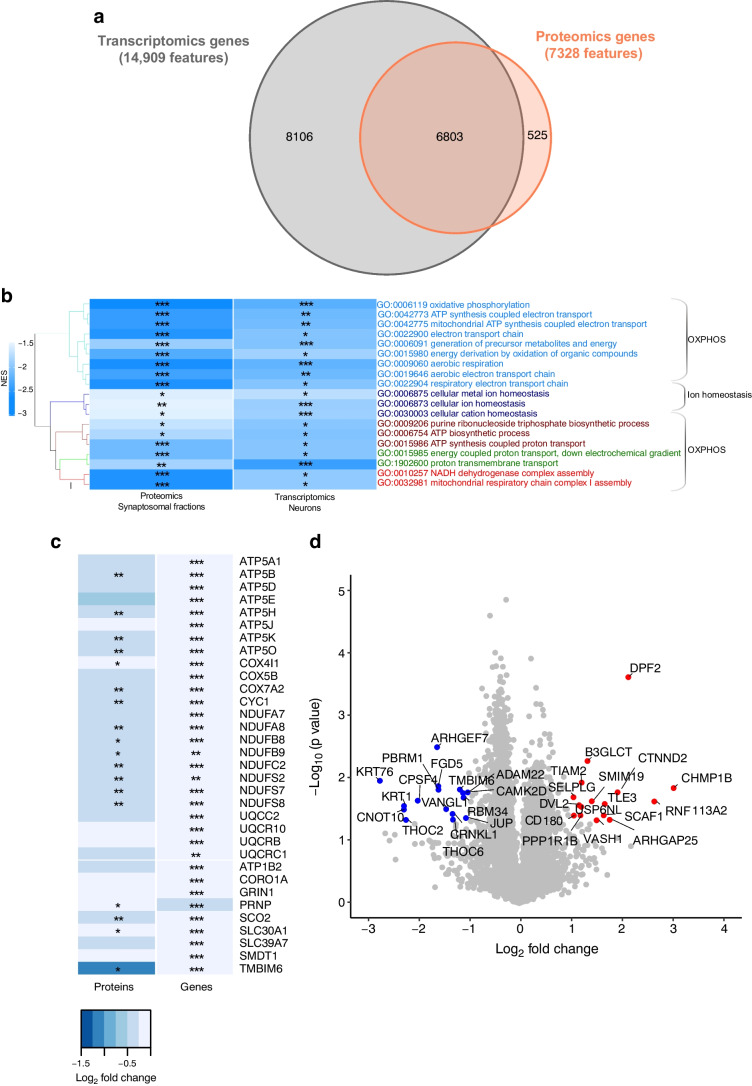
Methods: High-fat-diet fed and low-dose streptozocin-treated C57BL/6N mice were exposed to one (acute hypoglycaemia [AH]) or multiple (recurrent hypoglycaemia [RH]) insulin-induced hypoglycaemic episodes and plasma glucagon levels were measured. Single-nuclei RNA-seq (snRNA-seq) data were obtained from the hypothalamus and cortex of mice exposed to AH and RH. Proteomic data were obtained from hypothalamic synaptosomal fractions.

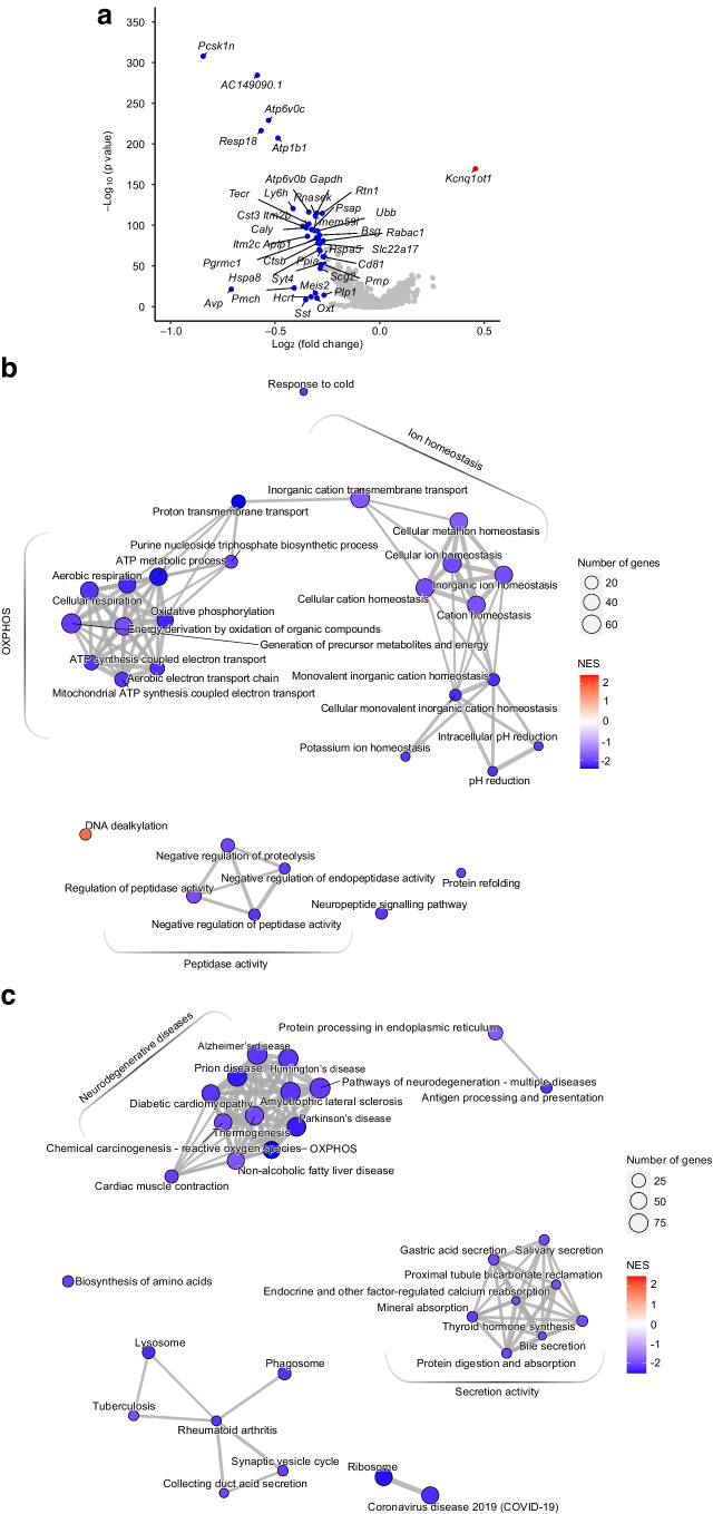
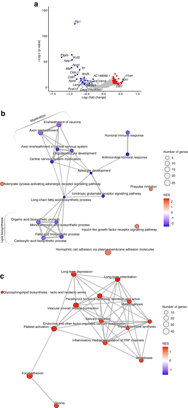
Results: The final insulin injection resulted in similar plasma glucose levels in the RH group and AH groups, but glucagon secretion was significantly lower in the RH group (AH: 94.5±9.2 ng/l [n=33]; RH: 59.0±4.8 ng/l [n=37]; p<0.001). Analysis of snRNA-seq data revealed similar proportions of hypothalamic cell subpopulations in the AH- and RH-exposed mice. Changes in transcriptional profiles were found in all cell types analysed. In neurons from RH-exposed mice, we observed a significant decrease in expression of Avp, Pmch and Pcsk1n, and the most overexpressed gene was Kcnq1ot1, as compared with AH-exposed mice. Gene ontology analysis of differentially expressed genes (DEGs) indicated a coordinated decrease in many oxidative phosphorylation genes and reduced expression of vacuolar H+- and Na+/K+-ATPases; these observations were in large part confirmed in the proteomic analysis of synaptosomal fractions. Compared with AH-exposed mice, oligodendrocytes from RH-exposed mice had major changes in gene expression that suggested reduced myelin formation. In astrocytes from RH-exposed mice, DEGs indicated reduced capacity for neurotransmitters scavenging in tripartite synapses as compared with astrocytes from AH-exposed mice. In addition, in neurons and astrocytes, multiple changes in gene expression suggested increased amyloid beta (Aβ) production and stability. The snRNA-seq analysis of the cortex showed that the adaptation to RH involved different biological processes from those seen in the hypothalamus.

Conclusions/interpretation: The present study provides a model of defective counterregulation in a mouse model of type 2 diabetes. It shows that repeated hypoglycaemic episodes induce multiple defects affecting all hypothalamic cell types and their interactions, indicative of impaired neuronal network signalling and dysegulated hypoglycaemia sensing, and displaying features of neurodegenerative diseases. It also shows that repeated hypoglycaemia leads to specific molecular adaptation in the hypothalamus when compared with the cortex.

Data availability: The transcriptomic dataset is available via the GEO ( http://www.ncbi.nlm.nih.gov/geo/ ), using the accession no. GSE226277. The proteomic dataset is available via the ProteomeXchange data repository ( http://www.proteomexchange.org ), using the accession no. PXD040183.

Keywords: Astrocytes; Counterregulation; Glucagon; Hypoglycaemia; Hypothalamus; Insulin; Neurodegeneration; Neurons; Oligodendrocytes; RNA-seq.


Figures

Fig. 1
Fig. 1
Recurrent exposure to hypoglycaemia reduces insulin-induced glucagon secretion. (a) Outline of the experiment to generate a model of type 2 diabetes and hypoglycaemia-associated autonomic failure (HAAF) in C57BL/6N male mice exposed to AH or RH. Created with BioRender.com. (b, c) Blood glucose (b) and plasma glucagon (c) in HFD-fed/STZ-treated mice exposed to AH or RH 1 h after a final i.p. injection of insulin (AH: n=33; RH: n=37). Data are means±SEM. ***p<0.001
Fig. 2
Fig. 2
Clustering and annotation of hypothalamic snRNA-seq in HFD-/STZ-treated mice exposed to RH and AH. (a) Two-dimensional Uniform Manifold Approximation and Projection (UMAP-1 and UMAP-2) representation of 14,979 and 14,934 nuclei isolated from the hypothalamus of AH- and RH-exposed mice, respectively. The cells are coloured based on the annotated cell type. (b) Violin plots of combined data from the RH and AH groups showing the expression of cellular-specific gene markers in each of the identified cell types. (c) Bar plot depicting the percentages of the different hypothalamic cell types per condition. Samples from n=3 mice were pooled for each group and the experiment was repeated once to obtain a biological replicate (n=2 integrated datasets per condition)
Fig. 3
Fig. 3
Transcriptional analysis of neurons from hypothalami of HFD-/STZ-treated mice exposed to AH or RH. Samples from n=3 mice were pooled for each group and the experiment was repeated once to obtain a biological replicate (n=2 integrated datasets per condition). (a) Volcano plot depicting differential expression of 14,909 genes from hypothalamic neurons of mice subjected to RH as compared with AH. The red dot outlines the upregulated DEG and blue dots outline downregulated DEGs (fold change >1.2 or <−1.2 and Bonferroni adjusted p value [padj]<0.05). (b, c) Network visualisation of the top enriched GO-BP (b) or KEGG (c) terms in DEGs in the RH vs AH group. Node colour indicates the normalised enrichment score (NES); node size indicates the number of core-enriched genes; edges (grey lines) represent the pairwise similarity between terms
Fig. 4
Fig. 4
Transcriptional analysis of oligodendrocytes from hypothalami of HFD-/STZ-treated mice exposed to AH or RH. Samples from n=3 mice were pooled for each group and the experiment was repeated once to obtain a biological replicate (n=2 integrated datasets per condition). (a) Volcano plot depicting differential expression of 10,814 genes from hypothalamic oligodendrocytes of mice subjected to RH as compared with AH. Red dots outline upregulated DEGs and blue dots outline downregulated DEGs (fold change >1.2 or <−1.2 and Bonferroni adjusted p value [padj]<0.05); only genes with log2 fold change >0.5 or l<−0.5 have been labelled. (b, c) Network visualisation of the enriched GO-BP (b) or KEGG (c) terms in DEGs in RH- vs AH-exposed mice. Node colour indicates the normalised enrichment score (NES); node size indicates the number of core-enriched genes overlapping gene count; edges (grey lines) represent the pairwise similarity between terms. TRP, transient receptor potential
Fig. 5
Fig. 5
Comparison of neuronal transcriptomic and synaptosomal proteomic data from HFD-/STZ-treated mice exposed to AH or RH. Proteomics data originated from the analysis of the hypothalami of n=4 mice/group. (a) Venn diagram illustrating the number of features analysed in the proteomics analysis of synaptosomal fractions and in the transcriptomics analysis of neuronal nuclei from the hypothalamus of mice exposed to RH vs AH. (b) Hierarchical clustering of GO-BP terms enriched in both synaptosomal fractions and neuron transcriptomes of hypothalamus from RH- vs AH-exposed mice (|normalised enrichment score [NES]| >1.5 and Benjamini–Hochberg adjusted p value [padj]<0.05). Colour of cells indicates the NES. Corresponding padj are also indicated (*p<0.05, **p<0.01, ***p<0.001). (c) Heatmap depicting log2 fold change in expression and p values (*p<0.05, **p<0.01, ***p<0.001) of selected features linked to OXPHOS or ion homeostasis. (d) Volcano plot depicting differential expression of 7328 proteins detected in hypothalamic synaptosomes from mice subjected to RH as compared with AH. Red and blue dots indicate proteins that are significantly differentially expressed (fold change >2 or <−2 and p<0.05). ATP1B2, sodium/potassium-transporting ATPase subunit beta-2; ATP5A1/ATP5B/ATP5D/ATP5E/ATP5H/ATP5K/ATP5O, mitochondrial ATP synthase subunit alpha/beta/delta/epsilon/d/e/O; ATP5J, ATP synthase-coupling factor 6, mitochondrial; CORO1A, Coronin-1A; GRIN1, glutamate receptor; COX4I1/COX5B/COX7A2, mitochondrial cytochrome c oxidase subunit 4 isoform 1/subunit 5B/subunit 7A2; CYC1, cytochrome c1, heme protein, mitochondrial; NDUFA7/NDUFA8, NADH dehydrogenase [ubiquinone] 1 alpha subcomplex subunit 8; NDUFB8, mitochondrial NADH dehydrogenase [ubiquinone] 1 beta subcomplex subunit 8; NDUFB9, NADH dehydrogenase [ubiquinone] 1 beta subcomplex subunit 9; NDUFC2, NADH dehydrogenase [ubiquinone] 1 subunit C2; NDUFS2/NDUFS7/NDUFS8, mitochondrial NADH dehydrogenase [ubiquinone] iron-sulfur protein 2/7/8; PRNP, major prion protein; SCO2, mitochondrial protein SCO2 homologue; SLC30A1, solute carrier family 30, member 1; SLC39A7, solute carrier family 39, member 7; SMDT1, single-pass membrane protein with aspartate rich tail 1; TMBIM6, Bax inhibitor motif containing 6; UQCC2, ubiquinol-cytochrome-c reductase complex assembly factor 2; UQCR10/UQCRB/QCRC1, cytochrome b-c1 complex subunit 9/7/1
Fig. 6
Fig. 6
Comparison of transcriptomic analysis from the hypothalamus and cortex of HFD-/STZ-treated mice exposed to AH or RH. For the hypothalamus, samples from n=3 mice were pooled for each group and the experiment was repeated once to obtain a biological replicate (n=2 integrated datasets per condition); a dataset of 14,979 nuclei from the AH group and 14,934 nuclei from the RH group was obtained. For the cortex, snRNA-seq analysis was conducted in duplicate using nuclei from the cortex of AH- and RH-exposed mice (samples from n=3 mice were pooled for each group) and a total of 4650 and 9088 nuclei were obtained, respectively. (a) Diagram representing the strategy followed. (b) Hierarchical clustering of GO-BP terms enriched in neuron transcriptomes (⎸normalised enrichment score [NES]⎹ >1.7) in the cortex and hypothalamus. Corresponding p values are also indicated (*p<0.05, **p<0.01, ***p<0.001)
Fig. 7
Fig. 7
Summary of the gene expression changes that may affect tripartite synapse function. In neurons, RH reduces the expression of OXPHOS genes (pink), of V- and Na+/K+-ATPase genes (brown), and of genes controlling neuropeptide expression, processing and synaptic vesicle exocytosis (violet). A decrease in OXPHOS-related gene expression reduces ATP production and, in combination with reduced expression of the Na+/K+-ATPase genes, overactivates GI neurons. In oligodendrocytes, many genes associated with myelination were downregulated, strongly suggesting a reduction in myelin formation capacity (yellow). In astrocytes, changes in gene expression suggest reduced neurotransmitter scavenging capacity (blue). In both neurons and astrocytes, several genes were downregulated that increased propensity for Aβ formation (green). NT, neurotransmitter. Created with BioRender.com

References

    1. Cryer PE. Hypoglycemia-associated autonomic failure in diabetes: maladaptive, adaptive, or both? Diabetes. 2015;64(7):2322–2323. doi: 10.2337/db15-0331. - DOI - PMC - PubMed
    1. MacDonald PE, Rorsman P. Metabolic messengers: glucagon. Nat Metab. 2023;5(2):186–192. doi: 10.1038/s42255-022-00725-3. - DOI - PubMed
    1. Thorens B. Neuronal regulation of glucagon secretion and gluconeogenesis. J Diabetes Investig. 2022;13(4):599–607. doi: 10.1111/jdi.13745. - DOI - PMC - PubMed
    1. Marty N, Dallaporta M, Thorens B. Brain glucose sensing, counterregulation, and energy homeostasis. Physiology. 2007;22:241–251. doi: 10.1152/physiol.00010.2007. - DOI - PubMed
    1. Stanley S, Moheet A, Seaquist ER. Central mechanisms of glucose sensing and counterregulation in defense of hypoglycemia. Endocr Rev. 2019;40(3):768–788. doi: 10.1210/er.2018-00226. - DOI - PMC - PubMed