

Endoscopic and histopathological hints on infections in patients of common variable immunodeficiency disorder with gastrointestinal symptoms
- PMID: 38017379
- PMCID: PMC10683160
- DOI: 10.1186/s12876-023-03052-3
Endoscopic and histopathological hints on infections in patients of common variable immunodeficiency disorder with gastrointestinal symptoms
Erratum in
-
Correction: Endoscopic and histopathological hints on infections in patients of common variable immunodeficiency disorder with gastrointestinal symptoms.BMC Gastroenterol. 2024 Jan 4;24(1):15. doi: 10.1186/s12876-023-03098-3. BMC Gastroenterol. 2024. PMID: 38177988 Free PMC article. No abstract available.
Abstract
Background and aims: Common variable immunodeficiency disorder (CVID) patients may have gastrointestinal (GI) involvement and suffer from infections, which are poorly understood. This study aimed to evaluate the clinical, endoscopic, and histopathological features of CVID patients with GI symptoms and determine their correlation with infections.
Methods: We performed a retrospective study on 21 CVID patients with GI symptoms who underwent endoscopic examination in Peking Union Medical College Hospital from 2000 to 2020. The clinical, infectious, endoscopic, and histopathological features were reassessed.
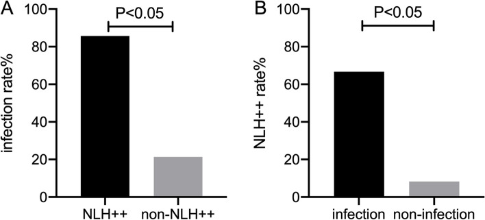
Results: Chronic diarrhea was the most prevalent GI symptom, observed in 95.2% of our CVID cohort. Over 85% of patients had low body weight and malabsorption. Small bowel villous atrophy was found in 90.5% of patients under endoscopy and mostly confirmed by histopathology. GI infections were identified in 9 (42.9%) patients. Of these, 7 patients with diffuse and obvious nodular lymphoid hyperplasia (NLH) of small bowel under endoscopy had significantly higher infection rate (85.7% vs 21.4%, p < 0.05), predominantly with Giardia and bacteria. Small bowel biopsies showed 95% of patients lacked plasma cells and 60% had increased intraepithelial lymphocytes (IELs), but not significantly different between GI infection and non-infection group. Most patients improved after intravenous immunoglobulin and anti-infection therapy.
Conclusions: CVID could involve GI tract, particularly small bowel. Obvious NLH under endoscopy could be a hint for GI infection in CVID patients. Comprehensive endoscopic and histopathological evaluation may be helpful in CVID diagnosis and identification of potential co-infection, leading to proper treatment.
Keywords: Common variable immunodeficiency disorder; Endoscopy; Histopathology; Infection; Nodular lymphoid hyperplasia.
© 2023. The Author(s).
Conflict of interest statement
The authors declare no competing interests.
Figures


References
MeSH terms
LinkOut - more resources
Full Text Sources
