. 2023 Nov 29;18(1):81.
doi: 10.1186/s13062-023-00425-2.

Long noncoding RNA KCNMA1-AS1 promotes osteogenic differentiation of human bone marrow mesenchymal stem cells by activating the SMAD9 signaling pathway

Affiliations

Long noncoding RNA KCNMA1-AS1 promotes osteogenic differentiation of human bone marrow mesenchymal stem cells by activating the SMAD9 signaling pathway

Zhaoyi Mai et al. Biol Direct. .

Abstract

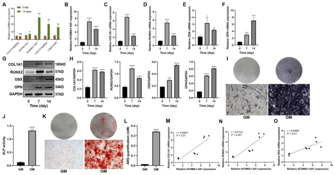
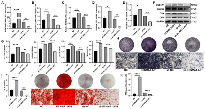
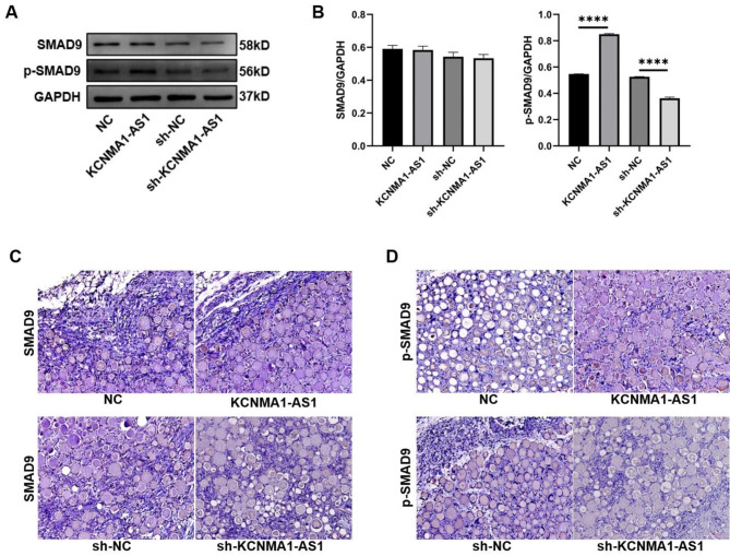
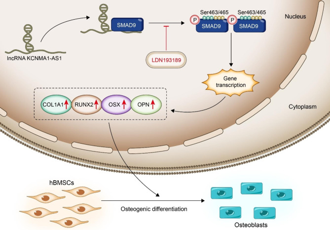
The human bone marrow mesenchymal stem cells (hBMSCs) undergo intense osteogenic differentiation, a crucial bone formation mechanism. Evidence from prior studies suggested an association between long noncoding RNAs (lncRNAs) and the osteogenic differentiation of hBMSCs. However, precise roles and molecular mechanisms are still largely unknown. In this work, we report for the first time that lncRNA KCNMA1 antisense RNA 1 (KCNMA1-AS1) plays a vital role in regulating hBMSCs' osteogenic differentiation. Here, it was observed that the KCNMA1-AS1 expression levels were significantly upregulated during osteogenic differentiation. In addition, KCNMA1-AS1 overexpression enhanced in vitro osteogenic differentiation of hBMSCs and in vivo bone formation, whereas knockdown of KCNMA1-AS1 resulted in the opposite result. Additionally, the interaction between KCNMA1-AS1 and mothers against decapentaplegic homolog 9 (SMAD9) was confirmed by an RNA pull-down experiment, mass spectrometry, and RIP assay. This interaction regulated the activation of the SMAD9 signaling pathway. Moreover, rescue assays demonstrated that the inhibitor of the SMAD9 signaling pathway reversed the stimulative effects on osteogenic differentiation of hBMSCs by KCNMA1-AS1 overexpression. Altogether, our results stipulate that KCNMA1-AS1 promotes osteogenic differentiation of hBMSCs via activating the SMAD9 signaling pathway and can serve as a biomarker and therapeutic target in treating bone defects.

Keywords: Human bone marrow mesenchymal stem cells; KCNMA1-AS1; Osteogenic differentiation; SMAD9; lncRNA.


Conflict of interest statement

The authors declare no competing interests.

Figures

Fig. 1
Fig. 1
KCNMA1-AS1 is upregulated when hBMSCs undergo osteogenic differentiation. A- Differentially expressed lncRNAs analyzed by qPCR after 14 days of osteogenic induction. B- Relative expression levels of KCNMA1-AS1 were measured using qPCR, and Glyceraldehyde 3-phosphate dehydrogenase (GAPDH) was used for normalization. C-F- Relative mRNA levels of COL1A1 (C), RUNX2 (D), OSX (E), and OPN (F) measured through qPCR, normalized to GAPDH. G and H- The protein levels of COL1A1, RUNX2, OSX, and OPN were detected by western blot. The internal reference is GAPDH. I and J- ALP staining (I) and ALP activity (J). hBMSCs cultured in osteogenic medium (OM) or growth medium (GM) for a week. Scale bar of microscopic images, 100 μm. K and L- ARS staining (K) and ARS quantification (L). hBMSCs were grown in an osteogenic medium (OM) or growth medium (GM) for two weeks. Scale bar of microscopical images, 100 μm. M-O- Correlation of the expression of KCNMA1-AS1 with that of COL1A1 (M), RUNX2 (N), and OSX (O) during osteogenic differentiation. ns: not significant, *P < 0.05, **P < 0.01, ***P < 0.001, ****P < 0.0001, compared with 0 day/GM.
Fig. 2
Fig. 2
KCNMA1-AS1 promotes osteogenic differentiation of hBMSCs in vitro. A- Transfection efficiency of KCNMA1-AS1 overexpression and KCNMA1-AS1 knockdown was measured by qPCR, normalized to GAPDH. B-E- Relative mRNA levels of COL1A1 (B), RUNX2 (C), OSX (D), and OPN (E) in hBMSCs transfected with lentivirus measured by qPCR after one week of osteogenic induction, normalized to GAPDH. F and G- The protein levels of COLA1, RUNX2, OSX, and OPN in hBMSCs transfected with lentivirus detected by western blot after one week of osteogenic induction. GAPDH was used as the internal reference. H and I ALP staining (H) and ALP activity (I) in hBMSCs transfected with lentivirus after one week of osteogenic induction. Scale bar of microscopical images, 100 μm. J and K- ARS staining (J) and ARS quantification (K) in hBMSCs transfected with lentivirus after two weeks of osteogenic induction. Scale bar of microscopical images, 100 μm. *P < 0.05, **P < 0.01, ***P < 0.001, ****P < 0.0001, in comparison to NC/sh-NC.
Fig. 3
Fig. 3
KCNMA1-AS1 promotes in vivo bone formation from hBMSCs. A- Xenograft tissue removed from the nude mice. B- Representative Micro CT scanning images of xenograft tissues. Scale bar, 500 μm. C-G- Bone volume or tissue volume (BV/TV) (C), bone surface/ tissue volume (BS/TV) (D), trabecular thickness (Tb.Th) (E), trabecular number (Tb.N) (F) and bone mineral density (BMD) (G) analyzed in xenograft tissues. H and I- HE staining (H) and Masson’s trichrome staining (I) of xenograft tissues. Scale bar, 50 μm. J-L- The expression levels of COL1A1 (J), RUNX2 (K), and OPN (L) in xenograft tissues evaluated by immunohistochemistry. Scale bar, 50 μm. *P < 0.05, **P < 0.01, in comparison to NC/sh-NC.
Fig. 4
Fig. 4
KCNMA1-AS1 directly binds to SMAD9 in hBMSCs. A and B- Subcellular localization of KCNMA1-AS1 in hBMSCs detected by the FISH assay. C- Mass spectrum of SMAD9. D- Colocalization of KCNMA1-AS1 and SMAD9 in hBMSCs detected by FISH assay and immunofluorescence. E- RIP assay analyzed by qPCR.
Fig. 5
Fig. 5
KCNMA1-AS1 activates the SMAD9 signaling pathway when hBMSCs undergo osteogenic differentiation. A-B- The SMAD9 protein levels and phosphorylated SMAD9 (p-SMAD9) protein levels in hBMSCs transfected with lentivirus detected by western blot after one week of osteogenic induction. GAPDH served as the reference. C-D- The expression of total SMAD9 (C) and p-SMAD9 (D) in xenograft tissues evaluated by immunohistochemistry. Scale bar, 50 μm. ****P < 0.0001, in comparison to NC/sh-NC.
Fig. 6
Fig. 6
Effect of KCNMA1-AS1 on osteogenic differentiation of hBMSCs with the SMAD9 signaling pathway activation. A and B- Protein levels of total SMAD9 and p-SMAD9 in lentivirus-transfected hBMSCs treated with DMSO or LDN193189 (100nM) detected by western blot after one week of osteogenic induction. GAPDH as the internal reference. C and D- The expression levels of COL1A1, RUNX2, OSX, and OPN in lentivirus-transfected hBMSCs treated with DMSO or LDN193189 (100nM) detected by western blot after one week of osteogenic induction. GAPDH as the reference. E and F- ALP staining (E) and ALP activity (F) in lentivirus-transfected hBMSCs treated with DMSO or LDN193189 (100nM) after one week of osteogenic induction. Scale bar of microscopical images, 100 μm. G and H- ARS staining (G) and ARS quantification (H) in lentivirus-transfected hBMSCs treated with DMSO or LDN193189 (100nM) after 14 days of osteogenic induction. Scale bar of microscopical images, 100 μm. ****P < 0.0001, compared with KCNMA1-AS1 + DMSO.
Fig. 7
Fig. 7
Schematic diagram illustrating the regulation of osteogenic differentiation in hBMSCs by KCNMA1-AS1.

References

    1. Tissue-engineered autologous. grafts for facial bone reconstruction | Science Translational Medicine [Internet]. [cited 2023 Sep 14]. Available from: https://www.science.org/doi/abs/10.1126/scitranslmed.aad5904. - DOI - PMC - PubMed
    1. Kinoshita Y, Maeda H. Recent developments of functional scaffolds for craniomaxillofacial bone tissue engineering applications. Sci World J. 2013;2013. - PMC - PubMed
    1. Cells | Free Full-Text. | Craniofacial Bone Tissue Engineering: Current Approaches and Potential Therapy [Internet]. [cited 2023 Sep 14]. Available from: https://www.mdpi.com/2073-4409/10/11/2993. - PMC - PubMed
    1. Grayson WL, Bunnell BA, Martin E, Frazier T, Hung BP, Gimble JM. Stromal cells and stem cells in clinical bone regeneration. Nat Rev Endocrinol. 2015;11(3):140–50. doi: 10.1038/nrendo.2014.234. - DOI - PMC - PubMed
    1. Park JJ, Rochlin DH, Parsaei Y, Shetye PR, Witek L, Leucht P et al. Bone tissue Engineering Strategies for Alveolar Cleft: review of preclinical results and guidelines for Future studies. Cleft Palate Craniofac J. 2022;10556656221104954. - PubMed

Publication types

MeSH terms

Substances

LinkOut - more resources