Review
. 2023 Nov 28;20(1):87.
doi: 10.1186/s12987-023-00489-2.

Breaking barriers: exploring mechanisms behind opening the blood-brain barrier

Affiliations
Review

Breaking barriers: exploring mechanisms behind opening the blood-brain barrier

Melanie E M Stamp et al. Fluids Barriers CNS. .

Abstract

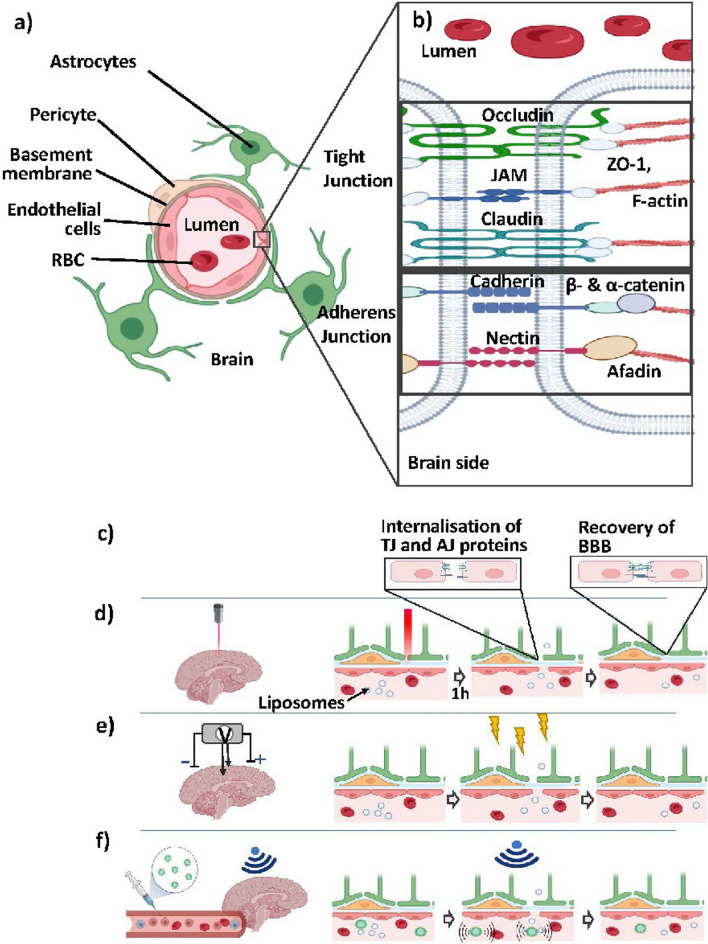
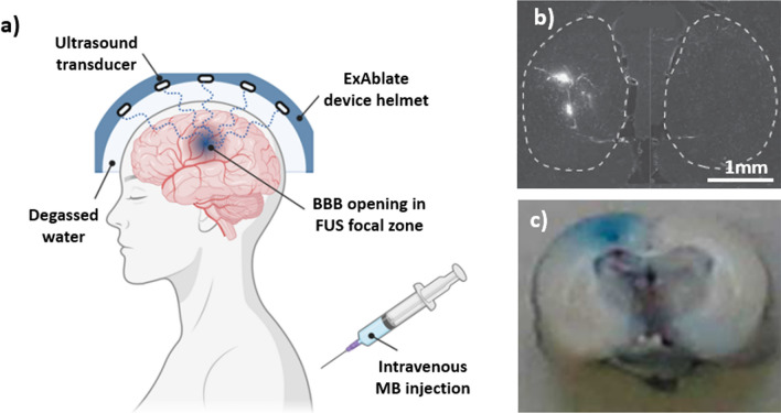
The blood-brain barrier (BBB) is a selectively permeable membrane that separates the bloodstream from the brain. While useful for protecting neural tissue from harmful substances, brain-related diseases are difficult to treat due to this barrier, as it also limits the efficacy of drug delivery. To address this, promising new approaches for enhancing drug delivery are based on disrupting the BBB using physical means, including optical/photothermal therapy, electrical stimulation, and acoustic/mechanical stimulation. These physical mechanisms can temporarily and locally open the BBB, allowing drugs and other substances to enter. Focused ultrasound is particularly promising, with the ability to focus energies to targeted, deep-brain regions. In this review, we examine recent advances in physical approaches for temporary BBB disruption, describing their underlying mechanisms as well as evaluating the utility of these physical approaches with regard to their potential risks and limitations. While these methods have demonstrated efficacy in disrupting the BBB, their safety, comparative efficacy, and practicality for clinical use remain an ongoing topic of research.

Keywords: BBB stimulation; Blood–Brain barrier; Focused ultrasound; Neurological therapeutics; Tight junctions.


Conflict of interest statement

The authors declare that they have no competing interests.

Figures

Fig. 1
Fig. 1
Schematic of a BBB. a Cell components. A basement membrane surrounds the endothelial cells that form the lumen. Surrounding these are pericytes and astrocytes, which together cover roughly 30 and 99% of the blood vessel, respectively. b Tight and adherens junctions. Apical and lumen TJs are composed of three transmembrane-spanning proteins: Occludin, Claudin, and JAM, which recruit ZO-1 (ZO-2, ZO-3), an actin-binding protein. Adjoining are AJs, which are composed of nectin- and cadherin-based adhesions. In the extracellular domain, nectins of neighbouring cells dimerise while the cytoplasmic tail recruits Afadin. The cadherin cytoplasmic tail recruits β-catenin, which binds to α-catinen that connects to the F-actin. c Schematics of BBB opening process during stimulation. TJ and AJ proteins internalise and retract to open the BBB for liposomes to trespass before recovery. d In photodynamic therapy, in which the brain is exposed to light for a certain time, post-exposure BBB opening, and subsequent recovery is observed. e Process in electroporation and non-invasive transcranial electrodes. During exposure to an electric field, TJ & AJ proteins functions reduce, allowing particles to pass paracellularly. Removal of stimulation leads to an almost instant BBB closure. f schematic of FUS with microbubbles injected intravenously prior to stimulation. During FUS stimulation, microbubbles within the capillaries are excited via external US, with vibrations opening the BBB, where removal of US leads to immediate closure of the BBB
Fig. 2
Fig. 2
a Schematic of photodynamic therapy-induced BBB opening in an animal model, adapted from Zhang et al. [68]. 5-ALA, a photosensitising agent, is injected intravenously prior to light exposure. Through a small opening/window in the skull, light at an agent-specific wavelength (here 635 nm) is emitted into the brain leading to photochemical reactions and heating of the stimulated area. The thermal effect leads to the local opening of the BBB post-exposure. b Schematic depiction of the LITT delivery system in mice. The laser fibre (right arrow) is positioned 1 mm caudal to the thermo-sensor (left arrow). In contrast to PD, a photosensitising agent is not used and laser treatment is delivered via laser fibre, avoiding a skull window. Figure adapted from Salehi et al. [87] c experimental results of PD stimulation: confocal imaging of GM1-liposomes the with usage of markers of the neurovascular unit. Cell nuclei are labelled with DAPI, liposome leakage outside the vascular endothelial cells are labelled by antibodies. The white arrows show the sites of liposome leakage. Images were taken from Zhang et al. [68]. d LITT increases BBB and BTB permeability in vivo. Representative white light and fluorescence images of mouse brains harvested on the indicated days after intravenous fluorescein injection. LITT was performed in the right somatosensory cortex. Control = unmanipulated brain. Scale bar = 5 mm, taken from Salehi et. al. [87]
Fig. 3
Fig. 3
Electrode arrangement for transcranial DC stimulation in a rat. a The epicranial electrode (contact area = 3.5 mm2) is fixed onto the skull unilaterally above the frontal cortex (1.5 mm right and 2 mm anterior to bregma) using dental cement. b Before DC stimulation, the epicranial electrode is filled with saline solution. A large rubber plate mounted on the chest serves as the counter electrode, images adapted from Liebetanz et al. [104]. c Schematic of the setup for the tDCS. One electrode connects to the rat cranium and the counter electrode connects to the ventral thoracic region, d Determination of the BBB solute permeability with illustration of the scanning region of interest (ROI) comprising several microvessels. The yellow frame enclosed area is the ROI used to determine the BBB permeability to a solute. e Total fluorescence intensity in the ROI as a function of the perfusion time. Fluorescence intensity in the figure is proportional to the total amount of solute accumulating in the region surrounding the microvessel. Figure taken from Shin et al. [101]
Fig. 4
Fig. 4
HFIRE stimulation, where a illustrates the distal end of the CETCS system with microneedles arborized from the primary cannula in convection-enhanced delivery CED catheters. b Schematic needle insertion in HFIRE induced BBB disruption and c post-contrast MRI images after gadolinium enhancement surrounding tumour area after treatment. Figures taken from Partridge et al. [119]. d MRI analysis of control brains in electroporation treatment with visible needle path (white arrow) obtained 30 min post treatment and e Histopathology of brain region with minimal bleeding along electrode insertion path. Figures taken from Sharabi et al. [61]. Immunofluorescent staining of transverse brain samples following intracranial HFIRE revealed a deceased in claudin-5 and ZO-1 reactivity 1 h post-treatment followed by gradual increase over time compared to control. Scale bar is 50 µm across all images. Image taken from Partridge et al. [118]
Fig. 5
Fig. 5
Schematic representation of blood–brain barrier opening in Alzheimer’s disease patient in vivo and patient-derived model in vitro. a Schematic of a magnetic resonance (MR)-guided ExAblate device used in the first successful blood–brain barrier (BBB) opening in Alzheimer’s disease (AD) patients. System consists of a hemispherical helmet lined with > 1000 independent transducer elements delivering low frequency ultrasound treatment to the prescribed target. The helmet is positioned in the specialised MRI bed with stereotaxic frame and the space between patient’s head and the helmet filled with degassed water for acoustic coupling. Microbubble (MB) administration is carried out using repeated bolus injection or a continuous infusion. Reversible BBB opening occurs in the defined ultrasound focal zone. Figure taken from [161]. b fluorescence images of FUS treated at US pressure of 0.84 MPa mouse brains (left side) compared to non-treated (right side), each injected with 500 kDa size dextran’s. Dashed lines indicated boundaries of the hippocampal regions. Figure taken from Chen et al. [142]. c Detection of BBB opening in rat brain, with left hemisphere sonicated at an intracranial pressure amplitude of 1.25 MPa in the presence of microbubbles at a dosage of 0.1 mL/kg and no treatment to the right side. Macroscopic inspection of 2–3 mm thick coronal section (front side of the brain) showed penetration of Evans Blue (molecular weight of 960.8 Da) into the underlying cortex. Figure taken from Alonso et al. [143]

References

    1. Abbott NJ. Inflammatory mediators and modulation of blood-brain barrier permeability. Cell Mol Neurobiol. 2000;20(2):131–147. doi: 10.1023/A:1007074420772. - DOI - PMC - PubMed
    1. Daneman R, Prat A. The blood-brain barrier. Cold Spring Harb Perspect Biol. 2015;7(1):a020412. doi: 10.1101/cshperspect.a020412. - DOI - PMC - PubMed
    1. Ballabh P, Braun A, Nedergaard M. The blood—brain barrier: an overview structure, regulation, and clinical implications. Neurobiol Dis. 2004;16:1–13. doi: 10.1016/j.nbd.2003.12.016. - DOI - PubMed
    1. Nelson AR, Sweeney MD, Sagare AP, Zlokovic BV. Neurovascular dysfunction and neurodegeneration in dementia and Alzheimer’s disease. Biochim Et Biophys Acta (BBA)—Mol Basis Dis. 2016;1862(5):887–900. doi: 10.1016/j.bbadis.2015.12.016. - DOI - PMC - PubMed
    1. Zhao Z, Nelson AR, Betsholtz C, Zlokovic BV. Perspective establishment and dysfunction of the blood-brain barrier. Cell. 2015;163(5):1064–1078. doi: 10.1016/j.cell.2015.10.067. - DOI - PMC - PubMed