Review
. 2023 Nov 29;18(1):89.
doi: 10.1186/s13024-023-00674-9.

Microglial function, INPP5D/SHIP1 signaling, and NLRP3 inflammasome activation: implications for Alzheimer's disease

Affiliations
Review

Microglial function, INPP5D/SHIP1 signaling, and NLRP3 inflammasome activation: implications for Alzheimer's disease

Gizem Terzioglu et al. Mol Neurodegener. .

Abstract

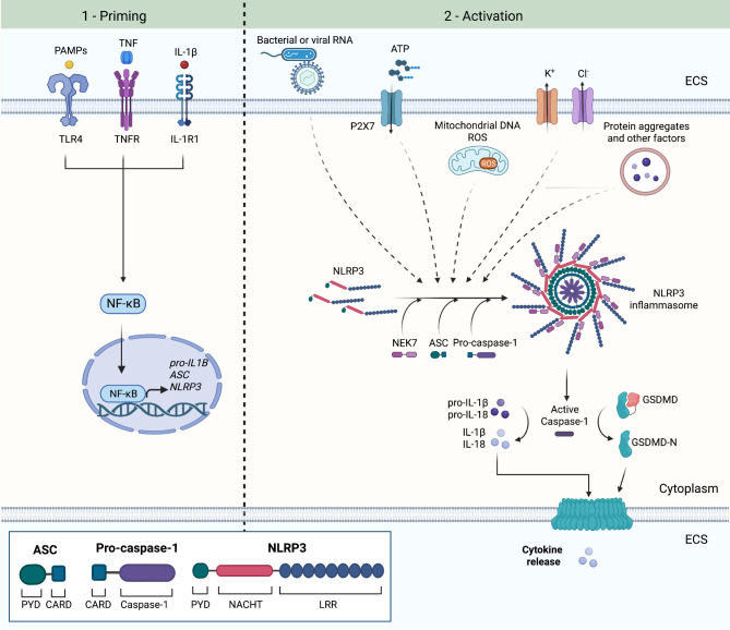
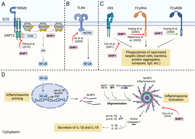
Recent genetic studies on Alzheimer's disease (AD) have brought microglia under the spotlight, as loci associated with AD risk are enriched in genes expressed in microglia. Several of these genes have been recognized for their central roles in microglial functions. Increasing evidence suggests that SHIP1, the protein encoded by the AD-associated gene INPP5D, is an important regulator of microglial phagocytosis and immune response. A recent study from our group identified SHIP1 as a negative regulator of the NLRP3 inflammasome in human iPSC-derived microglial cells (iMGs). In addition, we found evidence for a connection between SHIP1 activity and inflammasome activation in the AD brain. The NLRP3 inflammasome is a multiprotein complex that induces the secretion of pro-inflammatory cytokines as part of innate immune responses against pathogens and endogenous damage signals. Previously published studies have suggested that the NLRP3 inflammasome is activated in AD and contributes to AD-related pathology. Here, we provide an overview of the current understanding of the microglial NLRP3 inflammasome in the context of AD-related inflammation. We then review the known intracellular functions of SHIP1, including its role in phosphoinositide signaling, interactions with microglial phagocytic receptors such as TREM2 and evidence for its intersection with NLRP3 inflammasome signaling. Through rigorous examination of the intricate connections between microglial signaling pathways across several experimental systems and postmortem analyses, the field will be better equipped to tailor newly emerging therapeutic strategies targeting microglia in neurodegenerative diseases.

Keywords: Alzheimer’s Disease; INPP5D; Inflammation; Microglia; NLRP3 inflammasome; Phosphoinositide signaling; SHIP1.


Conflict of interest statement

The authors declare no competing interests.

Figures

Fig. 1
Fig. 1
NLRP3 inflammasome activation involves two steps. In the priming step, activation of TLR4, TNFR or IL-1R1 leads to activation of NF-kB, which subsequently translocates to the nucleus and induces the transcription of pro-IL-1β, ASC (also known as PYCARD) and NLRP3. NLRP3 is autoinihibted in the cytosol and remains in the autoinhibited state after the priming step. An activating signal is necessary to release the autoinhibition. Activated NLRP3 can undergo oligomerization through homotypic NACHT domain interactions to form the inflammasome complex. NLRP3 oligomers induce the filament assembly of ASC (PYCARD) into ASC specks through homotypic PYD interactions. ASC specks act as a molecular platform to recruit pro-caspase-1 through homotypic CARD interactions. Pro-caspase-1 undergoes autoproteolysis to generate active caspase-1, which can cleave pro-forms of IL-1β and IL-18 into their mature forms. Caspase-1 can also cleave gasdermin D (GSDMD) into its N-terminal fragment (GSDMD-N), which translocates to the plasma membrane and forms a pore, through which IL-1β and IL-18 are released into the extracellular space (ECS). NLRP3, nucleotide-binding domain (NOD)-like receptor protein 3; ASC, apoptosis-associated speck-like protein; PYD, pyrin-like domain; CARD, caspase recruitment domain; LRR, leucine-rich repeat. Figure created in BioRender.com [55, 61, 62]
Fig. 2
Fig. 2
SHIP1 is a phosphatase involved in phosphoinositide signaling. Activation of a receptor tyrosine kinase (RTK) recruits the p85 subunit of phosphotidylinositol-3 kinase (PI3K). PI3K generates PI(3,4,5)P3 at the plasma membrane by phosphorylating PI(4,5)P2. SYNJ1 can dephosphorylate PI(4,5)P2 back into its precursor, PI4P. PI(3,4,5)P3 can activate Akt, which can activate various downstream targets, one of which is IKKα of the IKK kinase complex. IKKα activation leads to phosphorylation of IkB, followed by the ubiquitin-mediated proteasomal degradation of IkB, which enables the NF-kB complex to translocate to the nucleus. The NF-kB complex can activate the transcription of its numerous target genes, including genes involved in immune response. SHIP1 can negatively regulate PI3K/Akt signaling. SHIP1 binds to PI(3,4,5)P3 via its PH-L domain and dephosphorylates PI(3,4,5)P3 via its 5’ phosphatase catalytic domain to generate PI(4,5)P2. C2 domain of SHIP1 can bind to PI(4,5)P2, which is thought to allosterically regulate SHIP1’s phosphatase activity. SH2 domain can dock onto phosphorylated tyrosine residues to regulate the activity of receptors containing immunoreceptor tyrosine activating/inhibiting motif (ITAMs/ITIMs). This domain is also important for SHIP1’s localization within the cell. SHIP1 can be phosphorylated at Ser440 within the 5’ phosphatase domain by PKA to increase the phosphatase activity. Phosphorylation of SHIP1 at Tyr1022 within the NPxY motif allows binding with SHIP1’s own SH2 domain, which may mediate its dimerization or oligomerization. ECS, extracellular space; SH2, src homology 2, PH-L, plectrin homology-like. Figure created in BioRender.com [87, 88, 91, 106, 107, 128]
Fig. 3
Fig. 3
SHIP1 regulates multiple receptors expressed in microglia and the microglial NLRP3 inflammasome. (A) TREM2 activation recruits p85 subunit of PI3K to the immunoreceptor tyrosine activating motif (ITAM) of DAP12. SHIP1 inhibits downstream activity of TREM2/DAP12 by binding to the immunoreceptor tyrosine activating motif (ITAM) of DAP12, preventing the association of p85 with DAP12, in osteoclasts. (B) SHIP1 inhibits the association of TLR4 with its adaptor protein MyD88 in macrophages. TLR4/MyD88 signaling leads to activation of NF-kB by inducing degradation of IΚB, which is also inhibited by SHIP1. (C) SHIP1 inhibits phagocytosis mediated by CR3 and FCγRIIA in macrophages. SHIP1 binds to the ITAM of FCγRIIA, inhibiting the downstream PI3K signaling. SHIP1 can also bind to the immunoreceptor tyrosine inhibitory motif (ITIM) of the inhibitory receptor FCγRIIB, which enhances FCγRIIB-mediated inhibition of phagocytosis. The exact mechanism of how SHIP1 inhibits CR3 is currently unknown. (D) SHIP1 inhibits the activation of the NLRP3 inflammasome. Pharmacological inhibition of SHIP1 or genetic reduction of INPP5D activates the NLRP3 inflammasome in human iPSC-derived microglial cells (iMGs), resulting in increased secretion of IL-1β and IL-18. On the other hand, loss of SHIP1 inhibits the upregulation of pro-IL-1β, suggesting that reduction of SHIP1 activity may induce inflammasome activation while inhibiting the priming step in some contexts. ECS, extracellular space. Figure created in BioRender.com [114, 121, 137, 138, 161, 197]

References

    1. Selkoe DJ. Alzheimer’s Disease: genes, proteins, and therapy. Physiol Rev. 2001;81:741–66. doi: 10.1152/physrev.2001.81.2.741. - DOI - PubMed
    1. Selkoe DJ, Hardy J. The amyloid hypothesis of Alzheimer’s Disease at 25 years. EMBO Mol Med. 2016;8:595. doi: 10.15252/emmm.201606210. - DOI - PMC - PubMed
    1. Sims R, van der Lee SJ, Naj AC, et al. Rare coding variants in PLCG2, ABI3, and TREM2 implicate microglial-mediated innate immunity in Alzheimer’s Disease. Nat Genet. 2017;49:1373–84. doi: 10.1038/ng.3916. - DOI - PMC - PubMed
    1. Dalmasso MC, Brusco LI, Olivar N, et al. Transethnic meta-analysis of rare coding variants in PLCG2, ABI3, and TREM2 supports their general contribution to Alzheimer’s Disease. Transl Psychiatry. 2019;9:1–6. doi: 10.1038/s41398-019-0394-9. - DOI - PMC - PubMed
    1. Efthymiou AG, Goate AM. Late onset Alzheimer’s Disease genetics implicates microglial pathways in Disease risk. Mol Neurodegener. 2017;12:1–12. doi: 10.1186/s13024-017-0184-x. - DOI - PMC - PubMed

MeSH terms

Substances