From teeth to pad: tooth loss and development of keratinous structures in sirenians
- PMID: 38018114
- PMCID: PMC10685118
- DOI: 10.1098/rspb.2023.1932
From teeth to pad: tooth loss and development of keratinous structures in sirenians
Abstract
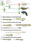
Sirenians are a well-known example of morphological adaptation to a shallow-water grazing diet characterized by a modified feeding apparatus and orofacial morphology. Such adaptations were accompanied by an anterior tooth reduction associated with the development of keratinized pads, the evolution of which remains elusive. Among sirenians, the recently extinct Steller's sea cow represents a special case for being completely toothless. Here, we used μ-CT scans of sirenian crania to understand how motor-sensor systems associated with tooth innervation responded to innovations such as keratinized pads and continuous dental replacement. In addition, we surveyed nine genes associated with dental reduction for signatures of loss of function. Our results reveal how patterns of innervation changed with modifications of the dental formula, especially continuous replacement in manatees. Both our morphological and genomic data show that dental development was not completely lost in the edentulous Steller's sea cows. By tracing the phylogenetic history of tooth innervation, we illustrate the role of development in promoting the innervation of keratinized pads, similar to the secondary use of dental canals for innervating neomorphic keratinized structures in other tetrapod groups.
Keywords: Steller's sea cow; dental pseudogenes; keratinous pad; sirenians; tooth loss.
Conflict of interest statement
We declare we have no competing interests.
Figures


Similar articles
-
Evolution of an extreme hemoglobin phenotype contributed to the sub-Arctic specialization of extinct Steller's sea cows.Elife. 2023 Jun 1;12:e85414. doi: 10.7554/eLife.85414. Elife. 2023. PMID: 37259901 Free PMC article.
-
Interordinal gene capture, the phylogenetic position of Steller's sea cow based on molecular and morphological data, and the macroevolutionary history of Sirenia.Mol Phylogenet Evol. 2015 Oct;91:178-93. doi: 10.1016/j.ympev.2015.05.022. Epub 2015 Jun 4. Mol Phylogenet Evol. 2015. PMID: 26050523
-
Genomic basis for skin phenotype and cold adaptation in the extinct Steller's sea cow.Sci Adv. 2022 Feb 4;8(5):eabl6496. doi: 10.1126/sciadv.abl6496. Epub 2022 Feb 4. Sci Adv. 2022. PMID: 35119923 Free PMC article.
-
Sirenian (manatees and dugongs) reproductive endocrinology.Gen Comp Endocrinol. 2024 Sep 15;356:114575. doi: 10.1016/j.ygcen.2024.114575. Epub 2024 Jun 20. Gen Comp Endocrinol. 2024. PMID: 38908455 Review.
-
Loss of teeth and enamel in tetrapods: fossil record, genetic data and morphological adaptations.J Anat. 2009 Apr;214(4):477-501. doi: 10.1111/j.1469-7580.2009.01060.x. J Anat. 2009. PMID: 19422426 Free PMC article. Review.
Cited by
-
Comparative genomics of sirenians reveals evolution of filaggrin and caspase-14 upon adaptation of the epidermis to aquatic life.Sci Rep. 2024 Apr 23;14(1):9278. doi: 10.1038/s41598-024-60099-2. Sci Rep. 2024. PMID: 38653760 Free PMC article.
-
How teeth, tusks and horny pads evolved together in sea cows.Proc Biol Sci. 2024 Aug;291(2028):20241154. doi: 10.1098/rspb.2024.1154. Epub 2024 Aug 14. Proc Biol Sci. 2024. PMID: 39137887 Free PMC article. No abstract available.
References
-
- Berkovitz B, Shellis P. 2018. The teeth of mammalian vertebrates. London, UK: Academic Press.
-
- Meredith RW, Zhang G, Gilbert MTP, Jarvis ED, Springer MS. 2014. Evidence for a single loss of mineralized teeth in the common avian ancestor. Science 346, 1254390. - PubMed
MeSH terms
Substances
LinkOut - more resources
Full Text Sources
Research Materials

