

Beraprost sodium attenuates the development of myocardial fibrosis after myocardial infarction by regulating GSK-3β expression in rats
- PMID: 38018586
- PMCID: PMC10633815
- DOI: 10.1002/iid3.1050
Beraprost sodium attenuates the development of myocardial fibrosis after myocardial infarction by regulating GSK-3β expression in rats
Abstract
Objective: The aim of this study was to elucidate the mechanism of beraprost sodium (BPS) in the intervention of myocardial fibrosis after myocardial infarction (MI) through glycogen synthase kinase-3β (GSK-3β) and to provide new ideas for intervention in myocardial fibrosis.
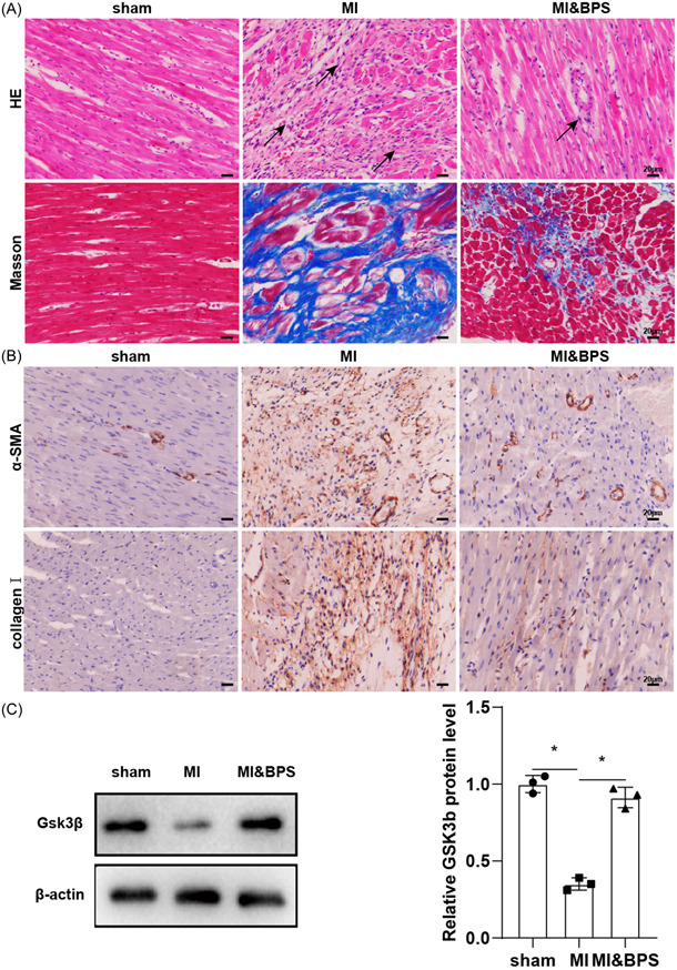
Materials and methods: MI model rats given BPS and cardiac fibroblasts (CFs) treated with BPS and TGF-β. HE staining and Masson staining were used to detect the pathological changes of myocardial tissue. Fibrotic markers were detected by immunohistochemical staining. The expressions of GSK-3β, cAMP response element binding protein (CREB), and p-CREB were analyzed by qPCR and western blot analysis. EDU staining was used to detect the proliferation of CFs. The promoter activity of GSK-3β was detected by luciferase assay. Chromatin immunoprecipitation assay was used to detect the binding levels of GSK-3β promoter and Y-box binding protein 1 (YBX1). The levels of intracellular cyclic adenosine monophosphate (cAMP) were analyzed by enzyme-linked immunosorbent assay (ELISA).
Results: After operation, BPS improved myocardial fibrosis and upregulated GSK-3β protein expression in male SD rats. BPS can down-regulate α-smooth muscle actin (α-SMA) level and up-regulate GSK-3β protein expression in CFs after TGF-β stimulation. Furthermore, GSK-3β knockdown can reverse the effect of BPS on TGF-β-activated CFs, enhance α-SMA expression, and promote the proliferation of CFs. BPS could regulate GSK-3β expression by promoting the binding of GSK-3β promoter to YBX1. BPS induced upregulation of p-CREB and cAMP, resulting in reduced fibrosis, which was reversed by the knockdown of GSK-3β or prostaglandin receptor (IPR) antagonists.
Conclusion: BPS treatment increased the binding of YBX1 to the GSK-3β promoter, and GSK-3β protein expression was upregulated, which further caused the upregulation of p-CREB and cAMP, and finally inhibited myocardial fibrosis.
Keywords: YBX1/GSK-3β; beraprost sodium; cAMP/p-CREB; myocardial fibrosis; myocardial infarction.
© 2023 The Authors. Immunity, Inflammation and Disease published by John Wiley & Sons Ltd.
Conflict of interest statement
The authors declare no conflict of interest.
Figures


Similar articles
-
[Mechanism of Linggui Zhugan Decoction in inhibiting myocardial fibrosis by regulating Wnt/β-catenin pathway].Zhongguo Zhong Yao Za Zhi. 2024 Aug;49(15):4178-4187. doi: 10.19540/j.cnki.cjcmm.20240415.701. Zhongguo Zhong Yao Za Zhi. 2024. PMID: 39307750 Chinese.
-
Qishen granule attenuates cardiac fibrosis by regulating TGF-β /Smad3 and GSK-3β pathway.Phytomedicine. 2019 Sep;62:152949. doi: 10.1016/j.phymed.2019.152949. Epub 2019 May 8. Phytomedicine. 2019. PMID: 31102891
-
[The mechanism of GSK-3β/CREB signaling pathway regulating macrophage pyroptosis and participating in the occurrence and development of diabetic foot ulcer].Xi Bao Yu Fen Zi Mian Yi Xue Za Zhi. 2024 Dec;40(12):1083-1088. Xi Bao Yu Fen Zi Mian Yi Xue Za Zhi. 2024. PMID: 39750046 Chinese.
-
MicroRNA-223 Regulates Cardiac Fibrosis After Myocardial Infarction by Targeting RASA1.Cell Physiol Biochem. 2018;46(4):1439-1454. doi: 10.1159/000489185. Epub 2018 Apr 19. Cell Physiol Biochem. 2018. PMID: 29689569
-
Entanglement of GSK-3β, β-catenin and TGF-β1 signaling network to regulate myocardial fibrosis.J Mol Cell Cardiol. 2017 Sep;110:109-120. doi: 10.1016/j.yjmcc.2017.07.011. Epub 2017 Jul 27. J Mol Cell Cardiol. 2017. PMID: 28756206 Free PMC article. Review.
References
-
- Yoshitomi T, Nagasaki Y. Self‐assembling antioxidants for ischemia‐reperfusion injuries. Antioxid Redox Signal. 2022;36(1‐3):70‐80. - PubMed
-
- Fan Q, Tao R, Zhang H, et al. Dectin‐1 contributes to myocardial ischemia/reperfusion injury by regulating macrophage polarization and neutrophil infiltration. Circulation. 2019;139(5):663‐678. - PubMed
-
- Ye L, D'Agostino G, Loo SJ, et al. Early regenerative capacity in the porcine heart. Circulation. 2018;138(24):2798‐2808. - PubMed
Publication types
MeSH terms
Substances
Grants and funding
LinkOut - more resources
Full Text Sources
Medical
Research Materials
