. 2024 Jun;20(6):1247-1269.
doi: 10.1080/15548627.2023.2288528. Epub 2023 Dec 5.

CARM1 drives mitophagy and autophagy flux during fasting-induced skeletal muscle atrophy

Affiliations

CARM1 drives mitophagy and autophagy flux during fasting-induced skeletal muscle atrophy

Derek W Stouth et al. Autophagy. 2024 Jun.

Abstract

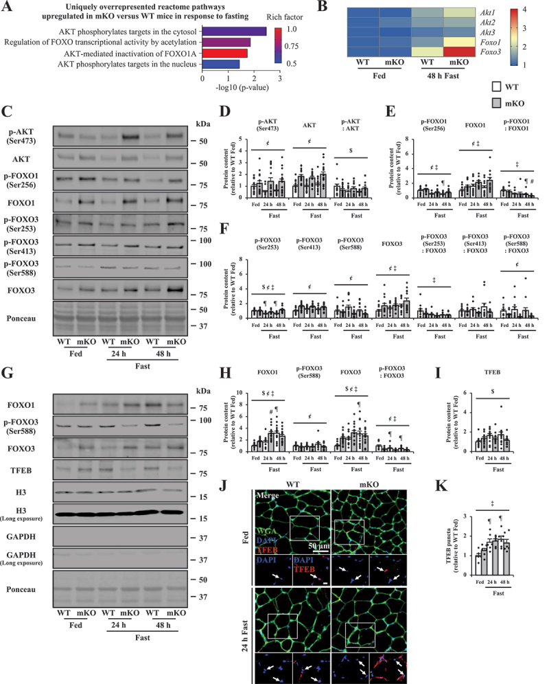
CARM1 (coactivator associated arginine methyltransferase 1) has recently emerged as a powerful regulator of skeletal muscle biology. However, the molecular mechanisms by which the methyltransferase remodels muscle remain to be fully understood. In this study, carm1 skeletal muscle-specific knockout (mKO) mice exhibited lower muscle mass with dysregulated macroautophagic/autophagic and atrophic signaling, including depressed AMP-activated protein kinase (AMPK) site-specific phosphorylation of ULK1 (unc-51 like autophagy activating kinase 1; Ser555) and FOXO3 (forkhead box O3; Ser588), as well as MTOR (mechanistic target of rapamycin kinase)-induced inhibition of ULK1 (Ser757), along with AKT/protein kinase B site-specific suppression of FOXO1 (Ser256) and FOXO3 (Ser253). In addition to lower mitophagy and autophagy flux in skeletal muscle, carm1 mKO led to increased mitochondrial PRKN/parkin accumulation, which suggests that CARM1 is required for basal mitochondrial turnover and autophagic clearance. carm1 deletion also elicited PPARGC1A (PPARG coactivator 1 alpha) activity and a slower, more oxidative muscle phenotype. As such, these carm1 mKO-evoked adaptations disrupted mitophagy and autophagy induction during food deprivation and collectively served to mitigate fasting-induced muscle atrophy. Furthermore, at the threshold of muscle atrophy during food deprivation experiments in humans, skeletal muscle CARM1 activity decreased similarly to our observations in mice, and was accompanied by site-specific activation of ULK1 (Ser757), highlighting the translational impact of the methyltransferase in human skeletal muscle. Taken together, our results indicate that CARM1 governs mitophagic, autophagic, and atrophic processes fundamental to the maintenance and remodeling of muscle mass. Targeting the enzyme may provide new therapeutic approaches for mitigating skeletal muscle atrophy.Abbreviation: ADMA: asymmetric dimethylarginine; AKT/protein kinase B: AKT serine/threonine kinase; AMPK: AMP-activated protein kinase; ATG: autophagy related; BECN1: beclin 1; BNIP3: BCL2 interacting protein 3; CARM1: coactivator associated arginine methyltransferase 1; Col: colchicine; CSA: cross-sectional area; CTNS: cystinosin, lysosomal cystine transporter; EDL: extensor digitorum longus; FBXO32/MAFbx: F-box protein 32; FOXO: forkhead box O; GAST: gastrocnemius; H2O2: hydrogen peroxide; IMF: intermyofibrillar; LAMP1: lysosomal associated membrane protein 1; MAP1LC3B: microtubule associated protein 1 light chain 3 beta; mKO: skeletal muscle-specific knockout; MMA: monomethylarginine; MTOR: mechanistic target of rapamycin kinase; MYH: myosin heavy chain; NFE2L2/NRF2: NFE2 like bZIP transcription factor 2; OXPHOS: oxidative phosphorylation; PABPC1/PABP1: poly(A) binding protein cytoplasmic 1; PPARGC1A/PGC-1α: PPARG coactivator 1 alpha; PRKN/parkin: parkin RBR E3 ubiquitin protein ligase; PRMT: protein arginine methyltransferase; Sal: saline; SDMA: symmetric dimethylarginine; SIRT1: sirtuin 1; SKP2: S-phase kinase associated protein 2; SMARCC1/BAF155: SWI/SNF related, matrix associated, actin dependent regulator of chromatin subfamily c member 1; SOL: soleus; SQSTM1/p62: sequestosome 1; SS: subsarcolemmal; TA: tibialis anterior; TFAM: transcription factor A, mitochondrial; TFEB: transcription factor EB; TOMM20: translocase of outer mitochondrial membrane 20; TRIM63/MuRF1: tripartite motif containing 63; ULK1: unc-51 like autophagy activating kinase 1; VPS11: VPS11 core subunit of CORVET and HOPS complexes; WT: wild-type.

Keywords: Atrophy; autophagy; coactivator-associated arginine methyltransferase 1; fasting; mitophagy; skeletal muscle.


Conflict of interest statement

No potential conflict of interest was reported by the authors.

Figures

Figure 1.
Figure 1.
Fasting-induced atrophy in carm1 mKO (skeletal muscle-specific knockout) mice. (A) Representative western blots of CARM1 protein content (normal and long exposures) in the TA (tibialis anterior) muscle from WT (wild-type) and mKO mice under fed and fast (24 and 48 h) conditions, as well as a representative Ponceau stain, below. Molecular masses (kDa) are shown at right of blots. (B) graphical summary of CARM1 protein expression in TA muscle of WT animals. Data are expressed as protein content relative to fed (n = 14). (C) body mass of WT and mKO animals in response to fasting (n = 11–21). (D) TA, quadricep (QUAD), and gastrocnemius (GAST) muscle mass from WT and mKO mice in fed and fast conditions. Muscle weight data are expressed relative to WT fed (n = 11–21). (E) Representative images of hematoxylin and eosin (H&E)-stained EDL muscle cross sections from WT and mKO mice in fed and fast cohorts. Scale bar: 50 μm. (F) graphical summary of the average myofiber cross-sectional area (CSA) of EDL muscles from WT and mKO mice in response to fasting. Data are expressed as CSA relative to the WT fed (n = 8–9). Data are means ± SEM. Two-way ANOVA; $ p < 0.05 interaction effect of genotype and fasting; ¢ p < 0.05 main effect of genotype; ‡ p < 0.05 main effect of fasting; ¶ p < 0.05 versus WT fed; # p < 0.05 versus mKO fed. (G) venn diagram representation of the number of significantly down- and upregulated genes in the TA muscle from WT and mKO mice based on RNA-seq following 48 h fast (n = 3). (H) Principal component (PC) analysis of transcript abundances in WT and mKO mice under fed and fast conditions (n = 3). (I) bubble plot of top 25 uniquely overrepresented gene ontology (GO) biological processes upregulated in mKO versus WT mice following 48 h fast (n = 3).
Figure 2.
Figure 2.
PRMT (protein arginine methyltransferase) content and activity in mKO animals following fasting-evoked muscle wasting. (A) heatmap of PRMT family members in TA muscle of WT and mKO mice after fasting. Data are expressed relative to WT fed (n = 3). (B) PRMT1, PRMT5, PRMT6, and PRMT7 mRNA expression in EDL muscles from WT and mKO mice during fed and fast conditions. Data are expressed as mRNA content relative to WT fed muscle (n = 9–13). (C) typical western blots of PRMT1, PRMT5, PRMT6, PRMT7, monomethylarginine (MMA), asymmetric dimethylarginine (ADMA), asymmetric arginine dimethylated CARM1 substrates, symmetric dimethylarginine (SDMA), asymmetrically dimethylated SMARCC1/BAF155 (SWI/SNF related, matrix associated, actin dependent regulator of chromatin, subfamily c, member 1) me2a (Arg1064), total SMARCC1, asymmetrically dimethylated PABPC1/PABP1 (poly(A) binding protein, cytoplasmic 1) me2a (Arg455/Arg460), and total PABPC1 in TA muscles from WT and mKO animals in fed and fast cohorts, accompanied by a typical Ponceau stain. Molecular masses (kDa) are shown at the right of blots. (D-G) graphical summaries of PRMT1, PRMT5, PRMT6, PRMT7, MMA, ADMA, CARM1 substrate, SDMA, SMARCC1 me2a (Arg1064), SMARCC1, SMARCC1 methylation status (i.e., the methylated form of the protein relative to the total amount of the protein), PABPC1 me2a (Arg455/Arg460), PABPC1, and PABPC1 methylation status in WT and mKO mice after fasting. Data are expressed as protein content relative to WT fed (n = 8–15). Data are means ± SEM. Two-way ANOVA; $ p < 0.05 interaction effect of genotype and fasting; ¢ p < 0.05 main effect of genotype; ‡ p < 0.05 main effect of fasting; ¶ p < 0.05 versus WT fed.
Figure 3.
Figure 3.
CARM1 regulates metabolic signaling during fasting-induced muscle atrophy. (A) Representative western blots of phosphorylated AMP-activated protein kinase (p-PRKAA/AMPK [Thr172]), total AMPK, PPARGC1A/PGC-1α (PPARG coactivator 1 alpha), CS (citrate synthase), and proteins indicative of mitochondrial OXPHOS (oxidative phosphorylation) complexes I – V (CI – CV) in WT and mKO TA muscles following fasting, accompanied by a typical Ponceau stain. Molecular masses (kDa) are shown at the right of blots. (B-C) graphical summaries of p-PRKAA/AMPK (Thr172), AMPK, AMPK phosphorylation status, PPARGC1A, CS, and total OXPHOS protein expression in WT and mKO TA muscles in response to fasting. Data are expressed as protein content relative to WT fed (n = 8–21). (D) Representative immunofluorescence images of MYH (myosin heavy chain) type I (blue), IIA (green), IIX (red), and IIB (black) in EDL muscles of WT and mKO mice after fasting. (E) graphical summaries of EDL myofiber MYH composition in both genotypes following food deprivation. (F) typical transmission electron micrographs of mitochondria and myonuclei (*) within the SS (subsarcolemmal) and IMF (intermyofibrillar) regions of TA muscle in WT and mKO mice under fed and 48 h fast conditions. Scale bar: 1 μm (n = 4). (G) Representative image from WT muscle of normal mitochondrial ultrastructure with intact membranes and clearly defined cristae (white arrow). Scale bar: 200 nm. (H) Transmission electron micrographs were analyzed for mitochondrial density (μm2 x # per μm2 x 100) within the SS and IMF areas of the muscle. (I) Representative images of succinate dehydrogenase (SDH)-stained EDL muscle cross-sections from WT and mKO mice after food deprivation. Scale bar: 50 μm (n = 5–6). (J) graphical summary of SDH staining intensity in both genotypes following fasting. (K-L) complex I-supported state III respiration and complex I + II-supported state III respiration in WT and mKO TA muscle under fed and 48 h fast conditions (n = 11–14). Data are means ± SEM. Two-way ANOVA; $ p < 0.05 interaction effect of genotype and fasting; ¢ p < 0.05 main effect of genotype; ‡ p < 0.05 main effect of fasting; ¶ p < 0.05 versus WT fed; # p < 0.05 versus mKO Fed.
Figure 4.
Figure 4.
Mitophagy in mKO mice during fasting-evoked muscle atrophy. (A) Representative western blots of MAP1LC3/LC3 (microtubule associated protein 1 light chain 3)-I, LC3-II, SQSTM1, TOMM20 (translocase of outer mitochondrial membrane 20), histone H3 (normal and long exposures), and GAPDH (glyceraldehyde-3-phosphate dehydrogenase; normal and long exposures) in mitochondria isolated from QUAD muscles of WT and mKO mice treated with Sal (saline) or Col (colchicine) under fed and 48 h fast conditions. Blots are accompanied by a typical Ponceau stain. Molecular masses (kDa) are shown at the right of blots. (B-C) graphical summaries of mitochondrial LC3-II and SQSTM1 flux in WT and mKO animals under fed and 48 h fast conditions. Data are expressed as protein content relative to WT fed (n = 12–14). (D) Representative image from mKO muscle of abnormal mitochondrial ultrastructure (black arrow) with disrupted membranes, loss of cristae, and vacuolization. (E) graphical summary of total number of abnormal mitochondria in WT and mKO mice treated with Sal or Col under fed and 48 h fast conditions. (F) Representative western blots of PRKN/parkin (parkin RBR E3 ubiquitin protein ligase), TOMM20, histone H3 (normal and long exposures), and GAPDH (normal and long exposures) in mitochondria isolated from QUAD muscles of WT and mKO animals following fasting. Blots are accompanied by a typical Ponceau stain and molecular masses (kDa) are shown at the right of blots. (G) graphical summary of western blot data comprising mitochondrial PRKN protein content in WT and mKO mice during fed and 48 h fast conditions. Data are expressed as protein content relative to WT fed (n = 10–13). (H) Representative images of EDL muscle cross sections stained for PRKN (red; scale bar: 2 μm) and TOMM20 (green; scale bar: 1 μm) with a merged image. (I) graphical summary of PRKN puncta in WT and mKO mice following food deprivation assessed by immunofluorescence. Data are expressed as number of PRKN puncta per 1,000 μm2 (n = 3). (J) Prkn, Bnip3 (BCL2 interacting protein 3) mRNA expression in EDL muscles from WT and mKO mice under fed and fast conditions. Data are expressed as mRNA content relative to WT fed (n = 11–13). (K) Representative western blots of PRKN and BNIP3 in WT and mKO TA muscles after fasting, accompanied by a typical Ponceau stain. Molecular masses (kDa) are shown at the right of blots. (L) graphical summary of PRKN and BNIP3 protein expression levels in WT and mKO animals during fed and fast conditions. Data are expressed as protein content relative to WT fed (n = 11–18). Data are means ± SEM. Two-way ANOVA; $ p < 0.05 interaction effect of genotype and fasting; ¢ p < 0.05 main effect of genotype; ‡ p < 0.05 main effect of fasting; § p < 0.05 main effect of Col; ¶ p < 0.05 versus WT fed; # p < 0.05 versus mKO Fed.
Figure 5.
Figure 5.
Skeletal muscle autophagy is dysregulated in mKO animals during atrophy. (A) Representative western blots of LC3-I, LC3-II, and SQSTM1 in TA muscles of WT and mKO mice treated with Sal or Col under fed and 48 h fast conditions, accompanied by a typical Ponceau stain. Molecular masses (kDa) are shown at the right of blots. (B-C) Graphical summaries of LC3-II and SQSTM1 flux in WT and mKO animals under fed and 48 h fast conditions. Data are expressed as protein content relative to WT fed (n = 12–16). (D) Representative image from mKO muscle of double-membrane autophagic vacuole (yellow arrow). Scale bar: 200 nm. (E) Graphical summary of total number of autophagic vacuoles in WT and mKO mice treated with Sal or Col under fed and 48 h fast conditions. (F) Heatmap of macroautophagy genes in TA muscle with enrichment unique to carm1 mKO after fasting. Data are expressed relative to WT fed (n = 3). (G) typical western blots of phosphorylated autophagy-related 16 (p-ATG16L1 [Ser278]), total ATG16L1, phosphorylated (p)-MTOR (mechanistic target of rapamycin kinase; Ser2448), total MTOR, p-ULK1 (unc-51 like autophagy activating kinase 1; Ser555), p-ULK1 (Ser757), total ULK1, TFEB (transcription factor EB), SKP2 (S-phase kinase associated protein 2), BECN1, LAMP1 (lysosomal associated membrane protein 1), and LAMP2 in WT and mKO TA muscles after food deprivation, accompanied by a Ponceau stain. Molecular masses (kDa) are shown at the right of blots. (H) Graphical summaries of p-ATG16L1 (Ser278), ATG16L1, ATG16L1 phosphorylation status. (I) Representative images of EDL muscle cross sections stained for laminin (aqua), DAPI (4’,6-diamidino-2-phenylindole dihydrochloride; blue), and p-ATG16L1 (Ser278; red). White arrows indicate p-ATG16L1 (Ser278) puncta. Scale bar of merged image: 25 μm (n = 3). (J-L) Graphical summaries of p-MTOR (Ser2448), MTOR, MTOR phosphorylation status, p-ULK1 (Ser555), p-ULK1 (Ser757), ULK1, ULK1 phosphorylation statuses, TFEB, SKP2, BECN1, LAMP1, and LAMP2 protein expression in WT and mKO animals following fasting. Data are expressed as protein content relative to WT fed (n = 9–18). Data are means ± SEM. Two-way ANOVA; $ p < 0.05 interaction effect of genotype and fasting; ¢ p < 0.05 main effect of genotype; ‡ p < 0.05 main effect of fasting; § p < 0.05 main effect of Col; ¶ p < 0.05 versus WT fed.
Figure 6.
Figure 6.
Altered atrophic signaling and myonuclear localization of catabolic proteins in mKO animals after fasting. (A) uniquely overrepresented upregulated reactome pathways in mKO versus WT mice following 48 h fast (n = 3). (B) heatmap of Akt1, Akt2, Akt3, Foxo1, and Foxo3 genes in TA muscle of WT and mKO mice after fasting. Data are expressed relative to WT fed (n = 3). (C) Representative western blots of p-AKT (Ser473), total AKT, p-FOXO1 (forkhead box O1; Ser256), total FOXO1, p-FOXO3 (Ser253), p-FOXO3 (Ser413), p-FOXO3 (Ser588), and total FOXO3 in WT and mKO TA muscles after food deprivation, accompanied by a typical Ponceau stain. Molecular masses (kDa) are shown at the right of blots. (D-F) graphical summaries of p-AKT (Ser473), AKT, AKT phosphorylation status, p-FOXO1 (Ser256), FOXO1, FOXO1 phosphorylation status, p-FOXO3 (Ser253), p-FOXO3 (Ser413), p-FOXO3 (Ser588), FOXO3, and FOXO3 phosphorylation statuses in WT and mKO animals in response to fasting. Data are expressed as protein content relative to WT fed (n = 10–18). (G) Representative western blots of total FOXO1, p-FOXO3 (Ser588), total FOXO3, TFEB, histone H3 (normal and long exposures), and GAPDH (normal and long exposures) in myonuclei isolated from WT and mKO GAST muscles following food deprivation, accompanied by a typical Ponceau stain. Molecular masses (kDa) are shown at the right of blots. (H-I) graphical summaries of myonuclear FOXO1, p-FOXO3 (Ser588), FOXO3, FOXO3 phosphorylation status, and TFEB protein content in WT and mKO mice after fasting. Data are expressed as nuclear protein content relative to WT fed (n = 12–15). (J) Representative images of EDL muscle cross sections stained for wheat germ agglutinin (WGA; green), DAPI (blue), and TFEB (red). Scale bar of merged image: 50 μm. Higher magnifications are inset with TFEB-positive myonuclei (white arrows). Scale bar of inset: 10 μm. (K) graphical summary of nuclear TFEB content in WT and mKO animals following food deprivation. Data are expressed as TFEB-positive myonuclei relative to WT fed (n = 6). Data are means ± SEM. Two-way ANOVA; $ p < 0.05 interaction effect of genotype and fasting; ¢ p < 0.05 main effect of genotype; ‡ p < 0.05 main effect of fasting; ¶ p < 0.05 versus WT fed; # p < 0.05 versus mKO Fed.
Figure 7.
Figure 7.
The impact of fasting on human skeletal muscle fiber size, CARM1 biology, and atrophy- and autophagy-related signaling. (A) Representative images of H&E-stained vastus lateralis QUAD muscle cross sections from healthy male participants collected 1 h postprandial (fed) and following 48 h of fasting (fast). Scale bar: 50 μm. (B) graphical summary of the average myofiber CSA of QUAD muscles from adult humans in response to fasting. Data are expressed as CSA relative to fed (n = 9). (C) distribution of CSA (μm2) in QUAD of fed (solid line) and fast (dashed line) human myofibers (n = 9). (D) heatmap visualizing the average mRNA expression of PRMT family members in QUAD muscle of adult humans during fed and 40 h fast conditions. Microarray data extracted from GSE28016 (n = 7). (E) Representative western blots of CARM1, asymmetric arginine dimethylated CARM1 substrates, SMARCC1 me2a (Arg1064), total SMARCC1, PABPC1 me2a (Arg455/Arg460), total PABPC1, PRKN, BNIP3, LC3-I, LC3-II, p-ATG16L1 (Ser278), total ATG16L1, p-ULK1 (Ser757), and total ULK1 in QUAD muscles from healthy humans in response to 48 h of fasting, accompanied by a typical Ponceau stain. Molecular masses (kDa) are shown at the right of blots. (F-N) graphical summaries of CARM1, CARM1 substrate, SMARCC1 me2a (Arg1064), SMARCC1, SMARCC1 methylation status, PABPC1 me2a (Arg455/Arg460), PABPC1, PABPC1 methylation status, PRKN, BNIP3, LC3-I, LC3-II, p-ATG16L1 (Ser278), ATG16L1, ATG16L1 phosphorylation status, p-ULK1 (Ser757), ULK1, and ULK1 phosphorylation status in human muscle after fasting. Data are expressed as protein content relative to fed (n = 6–7). Data are means ± SEM. Paired t-tests; * p < 0.05 versus fed.

References

    1. Bedford MT, Clarke SG.. Protein arginine methylation in mammals: who, what, and why. Mol Cell. 2009 Jan 16;33(1):1–13. PMID: 19150423; PMCID: PMC3372459. doi: 10.1016/j.molcel.2008.12.013. - DOI - PMC - PubMed
    1. Yang Y, Bedford MT. Protein arginine methyltransferases and cancer. Nat Rev Cancer. 2013. Jan;13(1):37–50. Epub 2012 Dec 13. PMID: 23235912. doi: 10.1038/nrc3409 - DOI - PubMed
    1. Fulton MD, Brown T, Zheng YG. The biological axis of protein arginine methylation and asymmetric dimethylarginine. Int J Mol Sci. 2019 Jul 6;20(13):3322. PMID: 31284549; PMCID: PMC6651691. 10.3390/ijms20133322 - DOI - PMC - PubMed
    1. Guccione E, Richard S. The regulation, functions and clinical relevance of arginine methylation. Nat Rev Mol Cell Biol. 2019. Oct;20(10):642–657. Epub 2019 Jul 26. Erratum in: Nat Rev Mol Cell Biol. 2019 Sep;20(9):567. PMID: 31350521. doi: 10.1038/s41580-019-0155-x. - DOI - PubMed
    1. Larsen SC, Sylvestersen KB, Mund A, et al. Proteome-wide analysis of arginine monomethylation reveals widespread occurrence in human cells. Sci Signal. 2016 Aug 30;9(443):rs9. PMID: 27577262. doi: 10.1126/scisignal.aaf7329. - DOI - PubMed

Publication types

Substances