Review
. 2023 Dec 20;87(4):e0018621.
doi: 10.1128/mmbr.00186-21. Epub 2023 Nov 29.

On the origin of the nucleus: a hypothesis

Affiliations
Review

On the origin of the nucleus: a hypothesis

Buzz Baum et al. Microbiol Mol Biol Rev. .

Abstract

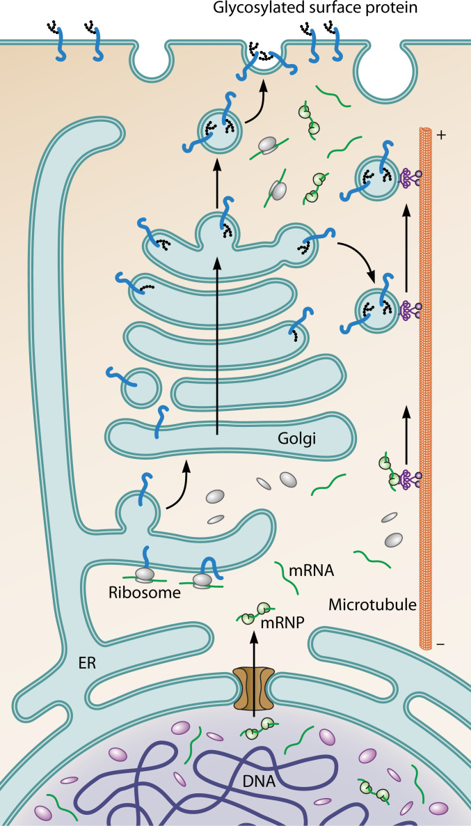
SUMMARYIn this hypothesis article, we explore the origin of the eukaryotic nucleus. In doing so, we first look afresh at the nature of this defining feature of the eukaryotic cell and its core functions-emphasizing the utility of seeing the eukaryotic nucleoplasm and cytoplasm as distinct regions of a common compartment. We then discuss recent progress in understanding the evolution of the eukaryotic cell from archaeal and bacterial ancestors, focusing on phylogenetic and experimental data which have revealed that many eukaryotic machines with nuclear activities have archaeal counterparts. In addition, we review the literature describing the cell biology of representatives of the TACK and Asgardarchaeaota - the closest known living archaeal relatives of eukaryotes. Finally, bringing these strands together, we propose a model for the archaeal origin of the nucleus that explains much of the current data, including predictions that can be used to put the model to the test.

Keywords: archaea; asgard archaea; eukaryogenesis; evolution; inside-out model; nucleolus; nucleus; origins; theory; topology.


Conflict of interest statement

The authors declare no conflict of interest.

Figures

Fig 1
Fig 1
These diagrams compare the spatial organization of gene expression in a schematic eukaryotic and bacterial cell. The images emphasize that, while transcription and translation are separate in eukaryotic cells, they are coupled in many bacteria. A key question posed by this article is whether or not transcription and translation are strongly coupled or partially uncoupled to enable the local translation of some transcripts in close archaeal relatives of eukaryotes.
Fig 2
Fig 2
Diagram shows the path of genetically encoded information in eukaryotic cells as it moves out of the nucleus (bottom) toward the cell periphery (top). DNA is transcribed in the nucleus. The RNAs generated are then processed and exported through nuclear pores into the cytoplasm. While many messenger RNAs (mRNAs) are rapidly translated upon entering the cytoplasm, others remain inaccessible to ribosomes as the result of RNA-binding proteins. Some of these are trafficked along microtubules in an inactive state, enabling local protein synthesis, e.g., at the tips of axons. In parallel, proteins carrying signal peptides, which are translated by ribosomes situated at the surface of the rough endoplasmic reticulum (ER), move through the ER and Golgi, where they are modified by glycosylation, packaged into vesicles, and trafficked out to the cell periphery along microtubules.
Fig 3
Fig 3
(A) Diagram shows the structure of the eukaryotic nucleus, and the endoplasmic reticulum to which it is connected. The nucleus is studded with nuclear pore complexes (NPCs). These NPCs sit at sites of high membrane curvature where the inner and outer nuclear membranes meet, and function as gated channels through which material can move between the nucleoplasm and cytoplasm. (B) Image shows a single eightfold symmetric nuclear pore complex inserted into the membrane viewed from one side (with kind permission of Agnieszka Obarska and Martin Beck). (C) New nuclear pores are inserted into the nuclear envelope via two processes: (i) insertion into gaps in the nuclear envelope as it reforms at mitotic exit and (ii) via interphase insertion. The diagram (adapted from Otsuke and Ellenberg, 2016, and based on electron microscopy data) shows a proposed path for interphase nuclear pore insertion from the inside out.
Fig 4
Fig 4
Schematic view of the tree of life with Bacteria in green, Archaea in purple, and eukaryotes (i.e. Eukaryota) in pink. Labels on the tree mark the Last Universal Common Ancestor (LUCA), the Last Archaeal Common Ancestor (LACA), the Last Bacterial Common Ancestor (LBCA), and the Last Eukaryotic Common Ancestor (LECA). As depicted in the diagram, LECA is hypothesized to be derived from the merging of at least two partners: an alphaproteobacterial symbiont and an asgardarchaeotal host.
Fig 5
Fig 5
Schematic tree of life with Bacteria in green, Archaea in purple, and Eukaryota in pink—emerging from the merger of an alphaproteobacterial symbiont with an asgardarchaeotal host. Horizontal gene transfer, which can complicate the phylogenetic analysis, is indicated by interconnecting lines. The light-shaded boxes indicate enzymes of likely archaeal (purple), bacterial (green), or unknown (gray) origin. Question marks indicate putative origins that are less clear.
Fig 6
Fig 6
Diagram depicts the stepwise evolution of the eukaryotic cell as imagined under the inside-out model (5). The cell in step 1 resembles a TACK archaeal cell. It possesses a nucleolar-like domain where rRNAs are assembled into ribosomes, a single bounding membrane, and a complete surface S-layer. The cell in step 2 has protrusions whose close contacts with bacterial partners (red) are facilitated by reduced S-layer coverage. The internal space within protrusions acts as a nascent cytoplasmic compartment, which is separated from the cell body by protrusion necks, where a region of high-membrane curvature is stabilized by multimeric proteins that bind to the membrane from the cytoplasmic side. These structures also function to confine the genome to the cell body. In step 3, the separation of a nascent nucleoplasm and cytoplasm is enhanced by the duplication of the machinery at protrusion necks (brown in the inset), and by the onset of directional, energy-dependent trafficking across this nuclear/cytoplasmic boundary. As a result, RNAs are only translated upon entry into the cytoplasm (indicated in green in inset). The increased curvature induced by duplication of the machinery at the neck of protrusions, together with the emergence of proteins that encourage the self-association of membranes, force the membrane to fold back over the cell body, effectively insulating the cell body from the chemical and physical environment. The partial fusion of protrusions leads to the formation of a more continuous cytoplasm. Proto-mitochondria reside in the spaces in between neighboring cytoplasmic compartments which are topologically equivalent to the lumen of the endoplasmic reticulum. Small black arrows indicate the flow of genetic information out from the center in steps 2 and 3. In the final step, step 4, the formation of a plasma membrane by a process of self-engulfment [as a single cytoplasmic bleb wraps around the whole and undergoes a single membrane scission event (see also Fig. 7)], yields a cell with a structure similar to that of a eukaryotic cell, with a topologically separate ER and plasma membrane, a continuous nuclear envelope-endoplasmic reticulum, and trapped vertically-inherited mitochondria, which later enter the cytoplasm. For an in-depth description of the entire process, see the original inside-out model.
Fig 7
Fig 7
Diagram shows an intermediate step in the process of eukaryogenesis expected under an outside-in model (left) and an inside-out model (right). Note that under the outside-in model, left, in order to generate a cell topologically similar to a eukaryotic cell (see Fig. 6, step 4), multiple membrane remodeling events are required to fuse initially separate endoplasmic reticulum (ER)-like compartments to generate a single lumenal network that is continuous with the nuclear envelope, and to generate a nuclear compartment that is connected to the cytoplasm via pores. In addition, additional membrane remodeling events are required to cut all the connections linking the nascent NE-ER compartment to the outer membrane to generate a topologically separate plasma membrane. By contrast, under the inside-out model shown in the diagram on the right, at this stage cells already possess a nascent nuclear compartment, a continuous ER and NE, and nuclear pores, but lack a single continuous cytoplasm and a plasma membrane. A topologically separate plasma membrane can be generated by a process of “auto-phagocytosis” whereby one protrusion extends around the whole and undergoes a single scission event.
Fig 8
Fig 8
This figure, kindly reproduced with permission of the authors from Rodrigues-Oliveira et al. (159), shows electron microscopic images of Lokiarchaeia cells. (A) An SEM shows a Candidatus Lokiarchaeum ossiferum cell (left) making contact with a possible syntropic partner via protrusions. (B) A zoomed in CryoEM image shows the highly curved neck that separates the Lokiarchaeum cell body from its protrusions, and the electron dense layer that underlies it. (C) and (D) show Candidatus Lokiarchaeum ossiferum cells in which actin filaments (ochre), ribosomes (gray), and the bounding membrane have been imaged using CryoEM and highlighted in different colors.

References

    1. Heimerl T, Flechsler J, Pickl C, Heinz V, Salecker B, Zweck J, Wanner G, Geimer S, Samson RY, Bell SD, Huber H, Wirth R, Wurch L, Podar M, Rachel R. 2017. A complex endomembrane system in the archaeon Ignicoccus hospitalis tapped by Nanoarchaeum equitans. Front Microbiol 8:1072. doi:10.3389/fmicb.2017.01072 - DOI - PMC - PubMed
    1. Greening C, Lithgow T. 2020. Formation and function of bacterial organelles. Nat Rev Microbiol 18:677–689. doi:10.1038/s41579-020-0413-0 - DOI - PubMed
    1. Volland J-M, Gonzalez-Rizzo S, Gros O, Tyml T, Ivanova N, Schulz F, Goudeau D, Elisabeth NH, Nath N, Udwary D, Malmstrom RR, Guidi-Rontani C, Bolte-Kluge S, Davies KM, Jean MR, Mansot J-L, Mouncey NJ, Angert ER, Woyke T, Date SV. 2022. A centimeter-long bacterium with DNA contained in metabolically active, membrane-bound organelles. Science 376:1453–1458. doi:10.1126/science.abb3634 - DOI - PubMed
    1. Katayama T, Nobu MK, Kusada H, Meng XY, Hosogi N, Uematsu K, Yoshioka H, Kamagata Y, Tamaki H. 2020. Isolation of a member of the candidate phylum 'Atribacteria' reveals a unique cell membrane structure. Nat Commun 11:6381. doi:10.1038/s41467-020-20149-5 - DOI - PMC - PubMed
    1. Baum DA, Baum B. 2014. An inside-out origin for the eukaryotic cell. BMC Biol 12:76. doi:10.1186/s12915-014-0076-2 - DOI - PMC - PubMed

LinkOut - more resources