. 2024 Jan 11;12(1):e0109023.
doi: 10.1128/spectrum.01090-23. Epub 2023 Nov 29.

Higher affinities of fibers with cell receptors increase the infection capacity and virulence of human adenovirus type 7 and type 55 compared to type 3

Affiliations

Higher affinities of fibers with cell receptors increase the infection capacity and virulence of human adenovirus type 7 and type 55 compared to type 3

Qiong Zhang et al. Microbiol Spectr. .

Abstract

HAdV-3, -7, and -55 are the predominant types causing acute respiratory disease outbreaks and can lead to severe and fatal pneumonia in children and adults. In recent years, emerging or re-emerging strains of HAdV-7 and HAdV-55 have caused multiple outbreaks globally in both civilian and military populations, drawing increased attention. Clinical studies have reported that HAdV-7 and HAdV-55 cause more severe pneumonia than HAdV-3. This study aimed to investigate the mechanisms explaining the higher severity of HAdV-7 and HAdV-55 infection compared to HAdV-3 infection. Our findings provided evidence linking the receptor-binding protein fiber to stronger infectivity of the strains mentioned above by comparing several fiber-chimeric or fiber-replaced adenoviruses. Our study improves our understanding of adenovirus infection and highlights potential implications, including in novel vector and vaccine development.

Keywords: adenoviruses; pathogenesis; pneumonia; receptors; respiratory viruses; virulence.


Conflict of interest statement

The authors declare no conflict of interest.

Figures

Fig 1
Fig 1
Infection and replication characterization of wild HAdV-3, HAdV-7, and HAdV-55. (a) Plaque formation of HAdV-3, HAdV-7, and HAdV-55 clinical isolates. Each symbol represents an individual isolate of the virus. (b) Viral genome of HAdV-3, HAdV-7, and HAdV-55. HAdVs in virus-infected A549 cells were determined at 4, 12, 24, 48, 72, and 96 h post-infection using qPCR. Plaque formation (c) and size distribution (d) of wild HAdV-3, HAdV-7, and HAdV-55. (e) A549 cells were infected with HAdV-3, HAdV-7, and HAdV-55 for 30, 60, or 240 min, washed three times, and the plaque formation units were detected. Each experiment was repeated three times independently, and the mean values and standard deviations were shown. Statistical analysis was performed using the Kruskal-Wallis test, followed by Dunn’s multiple comparisons test. ****P < 0.0001; ***P < 0.001.
Fig 2
Fig 2
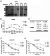
Affinity analysis of receptor DSG2 binding fiber knobs of HAdV-3, HAdV-7, and HAdV-55. (a) SDS-PAGE of the purified recombinant fiber knobs of HAdV-3, HAdV-7, HAdV-55, and HAdV-5. Purified protein in loading buffer was incubated at room temperature for 5 min and then incubated on ice (native, N) or denatured at 98°C for 5 min (boiled, B). The image labeled “Knob: Ad3” is identical to the image labeled “HAdV-B3-Knob” in our previously published article (37). The Ad3-knob protein used in both articles is the same one prepared from the same batch. (b) Comparison of the affinities of knobs binding DSG2 by ELISA. The plates were coated with 1.5 µg/mL DSG2 in HBS-N (Ca2+) and then incubated with serially diluted Ad3K, Ad7K, Ad55K, and Ad5K. Afterward, the HRP-labeled anti-His antibody was added. Finally, the substrate was added to read A450. (c) Affinity measurement and comparison of receptor DSG2 binding with fiber knobs of HAdV-3, HAdV-7, and HAdV-55 by SPR. (d and e) Knob competition experiments. Serially diluted recombinant knobs in phosphate-buffered saline (PBS) were added to A549 cells and incubated on ice. Subsequently, EGFP-expression HAdV rAd3EGFP (d) or rAd55EGFP (e) was added. After washing twice with cold PBS, the cells were cultured in a fresh medium for 2 days. Cells were photographed, and the number of fluorescent cells was determined. HAdV-5 fiber knob was used as the negative control.
Fig 3
Fig 3
Sketch of recombinant replication competent and fiber-chimeric rAd3E-K7 and rAdV3E-K55 preparation. rAd3E-K7 and rAd3E-K55 possess the same skeleton as rAd3E and a chimeric fiber with a replaced knob from HAdV-7 (green marked) and HAdV-55 (purple marked), respectively.
Fig 4
Fig 4
Infection and replication characterization of recombinant rAd3E, rAd3E-K7, and rAd3E-K55. Plaque size distribution (a) and plaque formation unit (b) of rAd3E, rAd3E-K7, and rAd3E-K55. (c) A549 cells were infected with rAd3E, rAd3E-K7, and rAd3E-K55 for 30 or 60 min, washed three times, and observed under a fluorescence microscope at 24 h post-infection. (d) Fluorescent cell numbers were counted 24 h post-infection. (e) Viral genome DNA proliferation curves and (f) infectious virus growth kinetics of rAd3E, rAd3E-K7, and rAd3E-K55. Viral genome copy numbers were determined by qPCR. Infectious virus titers were determined by counting fluorescence-forming units. The viruses infected in A549 cells were collected and determined at 4, 12, 24, 48, 72, and 96 h post-infection. Each experiment was repeated three times independently, and the mean values and standard deviations were shown. Statistical analysis was performed using the Kruskal-Wallis test, followed by Dunn’s multiple comparisons tests. P < 0.001; P < 0.01; P < 0.05.
Fig 5
Fig 5
Infection and replication characterization of recombinant rAd5-F3, rAd5-F7, and rAd5-F55. (a) Sketch of recombinant replication-deficient and fiber-replaced chimeric adenovirus type 5. rAd5-F3, rAd5-F7, and rAd5-F55 possess the same skeleton from rAd5 and replaced fiber from HAdV-3 (red marked), HAdV-7 (green marked), and HAdV-55 (purple marked), respectively. (b) Viral genome DNA proliferation curve kinetics of rAd5-F3, rAd5-F7, and rAd5-F55. Viral genome copy numbers were determined using qPCR. The viral particles in infected 293T cells were collected and measured at 4, 12, 24, 48, 72, and 96 h post-infection. (c) Plaque size distribution of rAd5-F3, rAd5-F7, and rAd5-F55. (d) 293T cells were infected with rAd5-F3, rAd5-F7, and rAd5-F55 for 30 or 60 min, washed three times, and observed under a fluorescence microscope at 24 h post-infection. Fluorescent cell numbers were counted. (e) Each experiment was repeated three times independently, and the mean values and standard deviations were shown. Statistical analysis was performed using the Kruskal-Wallis test, followed by Dunn’s multiple comparisons tests. P < 0.0001; P < 0.01; P < 0.05.
Fig 6
Fig 6
Infection and replication characterization of rAdV3E, rAdV7E, rAdV55E, rAdV3E-K7, and rAdV3E-K55 in HBEpiCs. (a) Infectious virus growth kinetics and (b) viral genome DNA proliferation curves of rAdV3E, rAdV7E, and rAdV55E. (c) Infectious virus growth kinetics and (d) viral genome DNA proliferation curves of rAdV3E, rAdV3E-K7, and rAdV3E-K55. Viral genome copy numbers were determined by qPCR. Infectious virus titers were determined by counting FFU. The viral particles in infected HBEpiCs were collected and quantified at 4, 12, 24, 48, 72, and 96 h post-infection. Each experiment was repeated three times independently, and the mean values and standard deviations were shown. Statistical analysis was performed using the Kruskal-Wallis test, followed by Dunn’s multiple comparisons tests. The two-way ANOVA with Dunnett’s multiple comarisons test was used to compare the means of groups at different times.
Fig 7
Fig 7
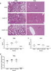
Humanized DSG2-KI mice intranasally infected with rAdV3E, rAdV3E-K7, and rAdV3E-K55. (a) Hematoxylin and eosin staining of lung and liver tissues. Arrows indicate pathological changes. (b) Lung injury scores and (c) liver injury scores. (d) Viral DNA load in lung tissues. Each symbol represents an individual mouse, and the horizontal lines indicate mean values or mean ± standard deviation. No significant differences were found (P > 0.05).

References

    1. Baker AT, Davies JA, Bates EA, Moses E, Mundy RM, Marlow G, Cole DK, Bliss CM, Rizkallah PJ, Parker AL. 2021. The fiber knob protein of human adenovirus type 49 mediates highly efficient and promiscuous infection of cancer cell lines using a novel cell entry mechanism. J Virol 95:e01849-20. doi:10.1128/JVI.01849-20 - DOI - PMC - PubMed
    1. Adams MJ, Lefkowitz EJ, King AMQ, Harrach B, Harrison RL, Knowles NJ, Kropinski AM, Krupovic M, Kuhn JH, Mushegian AR, Nibert M, Sabanadzovic S, Sanfaçon H, Siddell SG, Simmonds P, Varsani A, Zerbini FM, Gorbalenya AE, Davison AJ. 2017. Changes to taxonomy and the international code of virus classification and nomenclature ratified by the international committee on taxonomy of viruses. Arch Virol 162:2505–2538. doi:10.1007/s00705-017-3358-5 - DOI - PubMed
    1. Lion T. 2014. Adenovirus infections in immunocompetent and immunocompromised patients. Clin Microbiol Rev 27:441–462. doi:10.1128/CMR.00116-13 - DOI - PMC - PubMed
    1. Cederwall S, Påhlman LI. 2020. Respiratory adenovirus infections in immunocompetent and immunocompromised adult patients. Epidemiol Infect 147:e328. doi:10.1017/S0950268819002176 - DOI - PMC - PubMed
    1. Liu T, Zhou Z, Tian X, Liu W, Xu D, Fan Y, Liao J, Gu S, Li X, Zhou R. 2018. A recombinant trivalent vaccine candidate against human adenovirus types 3, 7, and 55. Vaccine 36:2199–2206. doi:10.1016/j.vaccine.2018.02.050 - DOI - PubMed