

A Three-Dimensional Trophoblast Invasion Microfluidic Platform for Toxicological Screening
- PMID: 38019404
- PMCID: PMC11138247
- DOI: 10.1007/978-1-0716-3495-0_18
A Three-Dimensional Trophoblast Invasion Microfluidic Platform for Toxicological Screening
Abstract
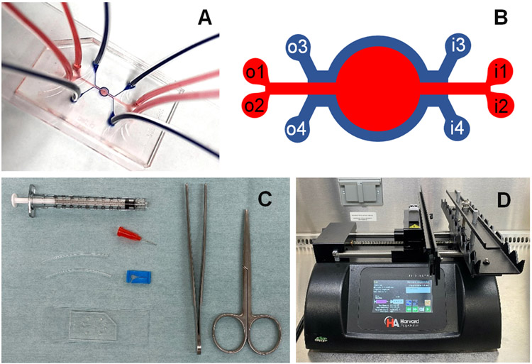
To improve our understanding of human placental function and placental cell responses to pregnancy stressors, the development of in vitro models that better recapitulate the in vivo placental microenvironment is needed. Here, we describe a three-dimensional (3D) silicone polymer polydimethylsiloxane (PDMS) microfluidic platform for modeling human trophoblast invasion recreating a placental heterocellular microenvironment. This platform allows the formation of a cellular barrier establishing a chemical gradient and real-time evaluation of trophoblast cell invasion and heterocellular cell-to-cell interactions.
Keywords: Invasion; Microfluidics; Three-dimensional; Trophoblast.
© 2024. The Author(s), under exclusive license to Springer Science+Business Media, LLC, part of Springer Nature.
Figures
Similar articles
-
A 3-dimensional microfluidic platform for modeling human extravillous trophoblast invasion and toxicological screening.Lab Chip. 2021 Feb 9;21(3):546-557. doi: 10.1039/d0lc01013h. Lab Chip. 2021. PMID: 33166377 Free PMC article.
-
A microfluidics assay to study invasion of human placental trophoblast cells.J R Soc Interface. 2017 May;14(130):20170131. doi: 10.1098/rsif.2017.0131. J R Soc Interface. 2017. PMID: 28566515 Free PMC article.
-
TGFβ signalling: a nexus between inflammation, placental health and preeclampsia throughout pregnancy.Hum Reprod Update. 2024 Jul 1;30(4):442-471. doi: 10.1093/humupd/dmae007. Hum Reprod Update. 2024. PMID: 38519450 Free PMC article. Review.
-
Placenta-on-a-chip: a novel platform to study the biology of the human placenta.J Matern Fetal Neonatal Med. 2016;29(7):1046-54. doi: 10.3109/14767058.2015.1038518. Epub 2015 Jun 15. J Matern Fetal Neonatal Med. 2016. PMID: 26075842 Free PMC article.
-
Investigation of human trophoblast invasion in vitro.Hum Reprod Update. 2020 Jun 18;26(4):501-513. doi: 10.1093/humupd/dmaa017. Hum Reprod Update. 2020. PMID: 32441309 Free PMC article. Review.
References
-
- Schmidt A, Morales-Prieto DM, Pastuschek J, Frohlich K, and Markert UR (2015) Only humans have human placentas: molecular differences between mice and humans. J Reprod Immunol 108, 65–71 - PubMed
-
- Barrientos G, Pussetto M, Rose M, Staff AC, Blois SM, and Toblli JE (2017) Defective trophoblast invasion underlies fetal growth restriction and preeclampsia-like symptoms in the stroke-prone spontaneously hypertensive rat. Mol Hum Reprod 23, 509–519 - PubMed
-
- Sebire NJ, Fox H, Backos M, Rai R, Paterson C, and Regan L (2002) Defective endovascular trophoblast invasion in primary antiphospholipid antibody syndrome-associated early pregnancy failure. Human reproduction 17, 1067–1071 - PubMed
-
- Liang CC, Park AY, and Guan JL (2007) In vitro scratch assay: a convenient and inexpensive method for analysis of cell migration in vitro. Nat Protoc 2, 329–333 - PubMed
MeSH terms
Substances
Grants and funding
LinkOut - more resources
Full Text Sources
