

Investigating the Spatiotemporal Summation of Perimetric Stimuli in Dry Age-Related Macular Degeneration
- PMID: 38019498
- PMCID: PMC10691387
- DOI: 10.1167/tvst.12.11.37
Investigating the Spatiotemporal Summation of Perimetric Stimuli in Dry Age-Related Macular Degeneration
Abstract
Purpose: To measure achromatic spatial, temporal, and spatiotemporal summation in dry age-related macular degeneration (AMD) compared to healthy controls under conditions of photopic gaze-contingent perimetry.
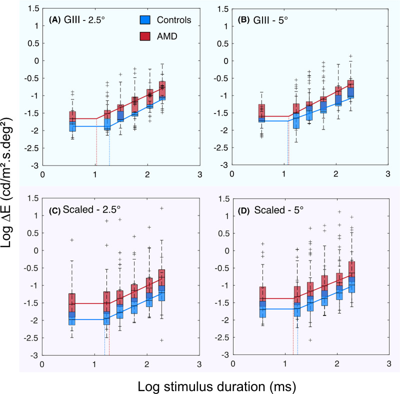
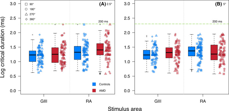
Methods: Twenty participants with dry AMD (mean age, 74.6 years) and 20 healthy controls (mean age, 67.8 years) performed custom, gaze-contingent perimetry tests. An area-modulation test generated localized estimates of Ricco's area (RA) at 2.5° and 5° eccentricities along the 0°, 90°, 180°, and 270° meridians. Contrast thresholds were measured at the same test locations for stimuli of six durations (3.7-190.4 ms) with a Goldmann III stimulus (GIII, 0.43°) and RA-scaled stimuli. The upper limit (critical duration) of complete temporal summation (using the GIII stimulus) and spatiotemporal summation (using the RA stimuli) was estimated using iterative two-phase regression analysis.
Results: Median (interquartile range [IQR]) RA estimates were significantly larger in AMD participants (2.5°: 0.21 [0.09-0.41] deg2; 5°: 0.32 [0.15-0.65 deg2]) compared to healthy controls (2.5°: 0.08 [0.05-0.13] deg2; 5°: 0.15 [0.08-0.22] deg2) at all test locations (all P < 0.05). No significant difference in median critical duration was found in AMD participants with the GIII stimulus (19.6 [9.9-30.4] ms) and RA-scaled stimuli (22.9 [13.9-40.3] ms) compared to healthy controls (GIII: 17.0 [11.3-24.0] ms; RA-scaled: 22.4 [14.3-33.1] ms) at all test locations (all P > 0.05).
Conclusions: Spatial summation is altered in dry AMD, without commensurate changes in temporal summation.
Translational relevance: The sensitivity of perimetry to AMD may be improved by utilizing stimuli that probe alterations in spatial summation in the disease.
Conflict of interest statement
Disclosure:
Figures


Similar articles
-
Spatiotemporal Summation of Perimetric Stimuli in Early Glaucoma.Invest Ophthalmol Vis Sci. 2015 Oct;56(11):6473-82. doi: 10.1167/iovs.15-16921. Invest Ophthalmol Vis Sci. 2015. PMID: 26447981
-
Investigating the linkage between mesopic spatial summation and variations in retinal ganglion cell density across the central visual field.Ophthalmic Physiol Opt. 2023 Sep;43(5):1179-1189. doi: 10.1111/opo.13158. Epub 2023 Apr 28. Ophthalmic Physiol Opt. 2023. PMID: 37118942
-
Temporal summation in myopia and its implications for the investigation of glaucoma.Ophthalmic Physiol Opt. 2023 Jul;43(4):788-797. doi: 10.1111/opo.13135. Epub 2023 Apr 3. Ophthalmic Physiol Opt. 2023. PMID: 37010917
-
A comparison of Goldmann III, V and spatially equated test stimuli in visual field testing: the importance of complete and partial spatial summation.Ophthalmic Physiol Opt. 2017 Mar;37(2):160-176. doi: 10.1111/opo.12355. Ophthalmic Physiol Opt. 2017. PMID: 28211185 Free PMC article.
-
Visual cycle modulators versus placebo or observation for the prevention and treatment of geographic atrophy due to age-related macular degeneration.Cochrane Database Syst Rev. 2020 Dec 17;12(12):CD013154. doi: 10.1002/14651858.CD013154.pub2. Cochrane Database Syst Rev. 2020. PMID: 33331670 Free PMC article.
References
-
- Flaxman SR, Bourne RRA, Resnikoff S, et al.. Global causes of blindness and distance vision impairment 1990-2020: a systematic review and meta-analysis. Lancet Glob Health. 2017; 5(12): e1221–e1234. - PubMed
-
- NICE. Age-Related Macular Degeneration. NICE Guideline [NG82]. Manchester, UK: National Institute for Health and Care Excellence; 2018. - PubMed
-
- Cheung CMG, Wong TY.. Treatment of age-related macular degeneration. Lancet. 2013; 382(9900): 1230–1232. - PubMed
MeSH terms
LinkOut - more resources
Full Text Sources
