. 2023 Dec 26;42(12):113503.
doi: 10.1016/j.celrep.2023.113503. Epub 2023 Nov 28.

Engineering CD276/B7-H3-targeted antibody-drug conjugates with enhanced cancer-eradicating capability

Affiliations

Engineering CD276/B7-H3-targeted antibody-drug conjugates with enhanced cancer-eradicating capability

Yang Feng et al. Cell Rep. .

Abstract

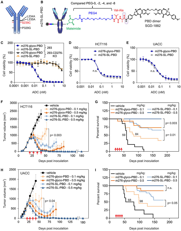
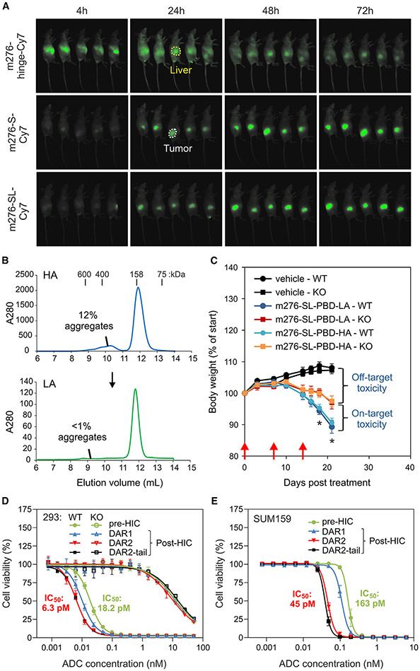
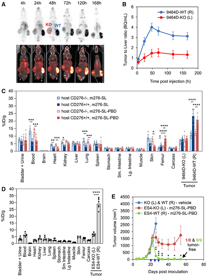
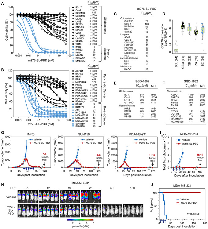
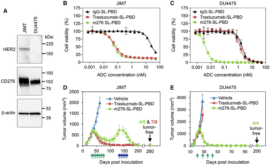
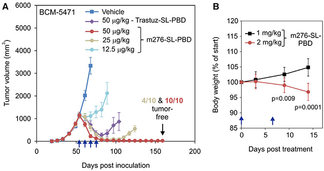
CD276/B7-H3 represents a promising target for cancer therapy based on widespread overexpression in both cancer cells and tumor-associated stroma. In previous preclinical studies, CD276 antibody-drug conjugates (ADCs) exploiting a talirine-type pyrrolobenzodiazepine (PBD) payload showed potent activity against various solid tumors but with a narrow therapeutic index and dosing regimen higher than that tolerated in clinical trials using other antibody-talirine conjugates. Here, we describe the development of a modified talirine PBD-based fully human CD276 ADC, called m276-SL-PBD, that is cross-species (human/mouse) reactive and can eradicate large 500-1,000-mm3 triple-negative breast cancer xenografts at doses 10- to 40-fold lower than the maximum tolerated dose. By combining CD276 targeting with judicious genetic and chemical ADC engineering, improved ADC purification, and payload sensitivity screening, these studies demonstrate that the therapeutic index of ADCs can be substantially increased, providing an advanced ADC development platform for potent and selective targeting of multiple solid tumor types.

Keywords: ADC; B7-H3; B7H3; CD276; CP: Cancer; PBD; TNBC; antibody-drug conjugate; breast cancer; m276-SL-PBD; pyrrolobenzodiazepine.


Conflict of interest statement

Declaration of interests Y.F., Z.Z., D.S.D., and B.S.C. are inventors of intellectual property related to m276 antibody-drug conjugates.

Figures

Figure 1.
Figure 1.. m276-SL-PBD structure and activity comparison with m276-glyco-PBD
(A) Amino acid substitutions in m276-SL. (B) Chemical structure of m276-SL-PBD linker and warhead: maleimide (green), PEG-4 spacer (blue), and cathepsin-B-cleavablevaline-alanine dipeptide (red). The gray cloud indicates the cleavable amide group. (C–E) Cell viability assays were used to measure the activity of m276-SL-PBD and m276-glyco-PBD against the parent 293 (CD276 wild type) or 293-CD276 KO (CD276 knockout) (C), HCT116 colon cancer (D), or UACC melanoma cells (E). Error bars denote SD. (F–I) Subcutaneous growth of HCT-116 (F) and UACC (H) tumors and corresponding Kaplan-Meier survival curves (HCT116, G; UACC, I). ADC treatments were initiated when tumors reached an average size of ~750 mm3 and were administered on the days shown (red arrows); n = 8–30/group; p values: t test (F and H) and log-rank test (G and I). Median survival is indicated for arms with <50% of animals alive at study end. Error bars denote SEM. n.s., non-significant.
Figure 2.
Figure 2.. Factors influencing m276-SL-PBD activity and toxicity
(A) In vivo fluorescence imaging of Cy7-labeled m276 antibodies in JIMT tumor-bearing mice at 4, 24, 48, and 72 h post injection. Side-view images of the tumor flank are shown. An example of tumor and liver fluorescence is highlighted (white and yellow regions of interest [ROIs], respectively). (B) SEC monitoring of m276-SL-PBD samples with high-aggregate (HA) and low-aggregate (LA) composition pre- and post purification by SEC. Size standards are shown at the top. (C) Body weights in CD276-WT and -KO mice after three treatments (red arrows) with 2 mg/kg of LA and HA ADC samples from (B). Student’s t test; *p < 0.05 for m276-SL-PBD-LA in WT versus KO and m276-SL-PBD-HA in WT versus KO; n = 8–15/group. (D and E) Cell viability assays measured the activity of m276-SL-PBD pre- and post-HIC purification against HEK293 CD276-WT, CD276-KO (D), or CD276+ SUM159 breast cancer cells (E). HIC-enriched DAR1, DAR2, and DAR2-tail fractions were tested (see Figure S5B). Error bars denote SD.
Figure 3.
Figure 3.. In vivo biodistribution of [89Zr]Zr-DFO-m276-SL and [89Zr]Zr-DFO-m276-SL-PBD
(A) PET imaging at ~4, 24, 48, 72, 120, and 168 h post injection of 0.5 mg/kg [89Zr]Zr-DFO-m276-SL-PBD ADC into mice with 9464D-CD276-WT (right flank) and 9464D-CD276-KO (left flank) tumors. (B) Quantification of tumor/liver labeling ratios from the PET study in (A). Error bars denote SD. (C) Biodistribution analysis 48 h post intravenous injection of [89Zr]Zr-DFO-m276-SL antibody or [89Zr] Zr-DFO-m276-SL-PBD ADC in CD276-wild-type (+/+) or -knockout (−/−) C57BL/6 mice. 9464D CD276-WT (right flank) and CD276-KO (left flank) tumors from the same mice were included for comparison. A one-way ANOVA determined p values between groups. Error bars denote SD. (D) Biodistribution analysis 48 h post injection of [89Zr]Zr-DFO-m276-SL-PBD ADC in athymic nude mice with ES4-CD276-WT (right flank, R) and ES4-CD276-KO (left flank, L) Ewing’s tumors. T test determined p values between WT and KO ES4 tumors. Error bars denote SD. (E) Subcutaneous growth of ES4-CD276-WT (right flank, R) or ES4-CD276-KO (left flank, L) Ewing’s tumors after m276-SL-PBD treatment. Treatments (blue arrows) were initiated when tumors reached an average size of ~750 mm3; n = 6–9/group. A t test determined p values between WT and KO ES4 tumors at the indicated time points. Error bars denote SEM. p values for (C)–(E): *p ≤ 0.05, **p ≤ 0.01, ***p ≤ 0.001, and ****p ≤ 0.0001.
Figure 4.
Figure 4.. Sensitivity to m276-SL-PBD across cancer types
(A and B) Cell viability assays measured m276-SL-PBD activity against glioblastoma/neuroblastoma (A) and pancreatic/breast cancer (B) cell lines. IC50 values are in the key. Error bars were omitted for clarity; SD was always <10%. (C) Table of IC50 values from cell viability assays showing the relative sensitivity of cancer cell lines to m276-SL-PBD. (D) CD276 mRNA expression in CD276-low B cell lymphocytic leukemia (BLL) and CD276-high glioblastoma (GBM), neuroblastoma (NB), pancreatic cancer (PC), and breast cancer (BC) cell lines using the database: DepMap mRNA expression. The number of cancer cell lines in each group is indicated in parentheses. Sensitive (blue dots) and resistant (back dot) cell lines used in the cell viability assays (A and B) are highlighted. (E and F) Tables showing glioblastoma/neuroblastoma (E) or pancreatic/breast cancer (F) cell line sensitivity (IC50 values from cell viability assays) after SGD-1882-free drug treatment and CD276 surface expression levels (mean fluorescence intensity; MFI) as measured by flow cytometry. (G) Subcutaneous (IMR5) and orthotopic (SUM159 and MDA-MB-231) tumor volumes following ADC treatment. Treatments with 0.5 mg/kg m276-SL-PBD (blue arrows) were initiated when tumors reached ~1,000 mm3; n = 8–10/group. Error bars denote SEM. (H) Bioluminescence imaging monitored systemic MDA-MB-231-luc tumor burden following intravenous injection into NRG mice, followed by randomization and treatment with vehicle or m276-SL-PBD. Representative mice shown (n = 15/group; DPI, days post inoculation). (I) Quantification of tumor burden from the MDA-MB-231 metastasis study in (H). Error bars denote SD. (J) Kaplan-Meier survival curves for the MDA-MB-231-luc metastases study in (H).
Figure 5.
Figure 5.. m276-SL-PBD targets CD276+ breast cancer independent of HER2 status
(A) Western blot analysis of CD276 and HER2 levels in JIMT and DU4475 breast cancer cells. (B and C) Cell viability assays measured trastuzumab-SL-PBD and m276-SL-PBD activity against JIMT (B) and DU4475 (C) breast cancer cells. Non-specific IgG-SL-PBD served as a non-binding control. Error bars denote SD. (D and E) Orthotopic growth of JIMT (D) and DU4475 (E) breast tumors in response to trastuzumab-SL-PBD and m276-SL-PBD treatment. ADC treatments (0.5 mg/kg) were initiated when tumor volumes reached ~750–1,000 mm3 and administered on the days shown (green arrows). Relapsed tumors in the m276-SL-PBD group were re-treated five times with ADC (blue arrows) when tumor volumes reached an average of ~750 mm3; n = 5–8/group. Error bars denote SEM.
Figure 6.
Figure 6.. Low-dose m276-SL-PBD induces durable tumor regression in multiple CD276+ breast cancer PDX models
(A and B) Immunofluorescence staining for CD276 (green) in BCM-5471, BCM-4013, and BCM-4272 (B) breast cancer PDX models. Tumor endothelium was co-stained with CD31 (red), confirming CD276 expression in both tumor cells and tumor vasculature. Non-specific IgG (A) served as a non-binding control. Scale bars in (A) and (B), 50 μm. (C–E) Orthotopic growth of BCM-5471 (C), BCM-4013 (D), and BCM-4272 (E) breast tumors following treatment with vehicle (control) or 50 or 100 μg/kg m276-SL-PBD. Treatments with m276-SL-PBD were initiated when tumor volumes reached ~1,000 mm3 and were administered on the days shown (blue arrows); n = 7–8/group. Error bars denote SEM.
Figure 7.
Figure 7.. Low m276-SL-PBD doses: Well tolerated with potent anti-tumor activity
(A) Orthotopic growth of BCM-5471 breast tumors in response to treatment with vehicle (control) or 12.5, 25, or 50 μg/kg m276-SL-PBD or 50 μg/kg trastuzumab-SL-PBD. ADC treatments were initiated when tumor volumes reached ~1,000 mm3 and were administered on the days shown (blue arrows); n = 10/group. p = 0.0003 between the 12.5 and the 25 μg/kg groups at day 89 post inoculation, and p = 0.001 between the 25 and the 50 μg/kg groups at day 124 post inoculation. Error bars denote SEM. (B) Body weights were evaluated in C57BL/6 mice following two treatments (blue arrows) with 1 or 2 mg/kg m276-SL-PBD. A t test determined p values between the two doses at the indicated time points. Error bars denote SD.

References

    1. Beck A, Goetsch L, Dumontet C, and Corvaïa N (2017). Strategies and challenges for the next generation of antibody-drug conjugates. Nat. Rev. Drug Discov 16, 315–337. - PubMed
    1. Amiri-Kordestani L, Blumenthal GM, Xu QC, Zhang L, Tang SW, Ha L, Weinberg WC, Chi B, Candau-Chacon R, Hughes P, et al. (2014). FDA approval: ado-trastuzumab emtansine for the treatment of patients with HER2-positive metastatic breast cancer. Clin. Cancer Res 20, 4436–4441. - PubMed
    1. Fuentes-Antrás J, Genta S, Vijenthira A, and Siu LL (2023). Antibody-drug conjugates: in search of partners of choice. Trends Cancer 9, 339–354. - PubMed
    1. Coats S, Williams M, Kebble B, Dixit R, Tseng L, Yao NS, Tice DA, and Soria JC (2019). Antibody-Drug Conjugates: Future Directions in Clinical and Translational Strategies to Improve the Therapeutic Index. Clin. Cancer Res 25, 5441–5448. - PubMed
    1. Criscitiello C, Morganti S, and Curigliano G (2021). Antibody-drug conjugates in solid tumors: a look into novel targets. J. Hematol. Oncol 14, 20. - PMC - PubMed

Publication types