. 2023 Dec;9(48):eadi9134.
doi: 10.1126/sciadv.adi9134. Epub 2023 Nov 29.

GDF8 inhibition enhances musculoskeletal recovery and mitigates posttraumatic osteoarthritis following joint injury

Affiliations

GDF8 inhibition enhances musculoskeletal recovery and mitigates posttraumatic osteoarthritis following joint injury

Camille R Brightwell et al. Sci Adv. 2023 Dec.

Abstract

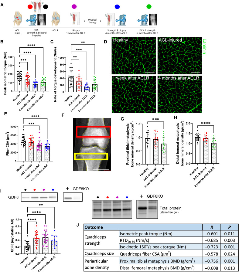
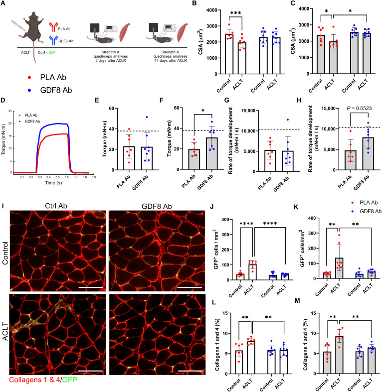
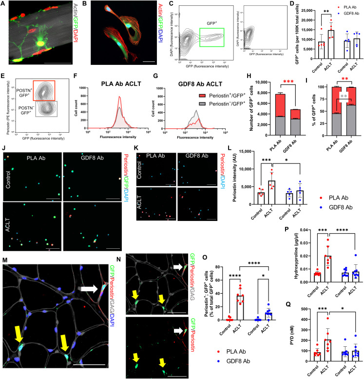
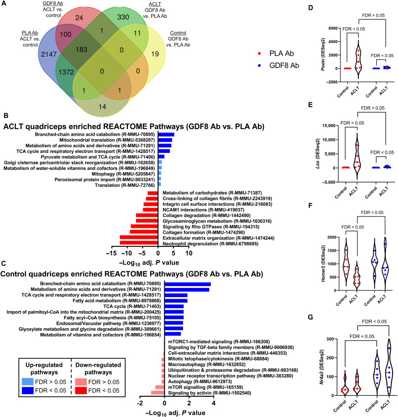
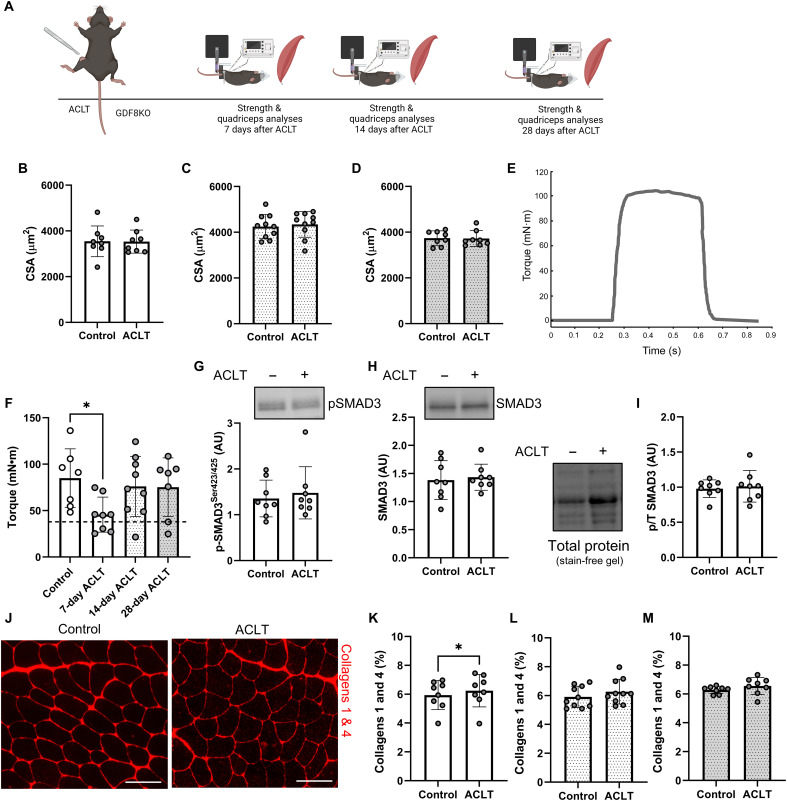
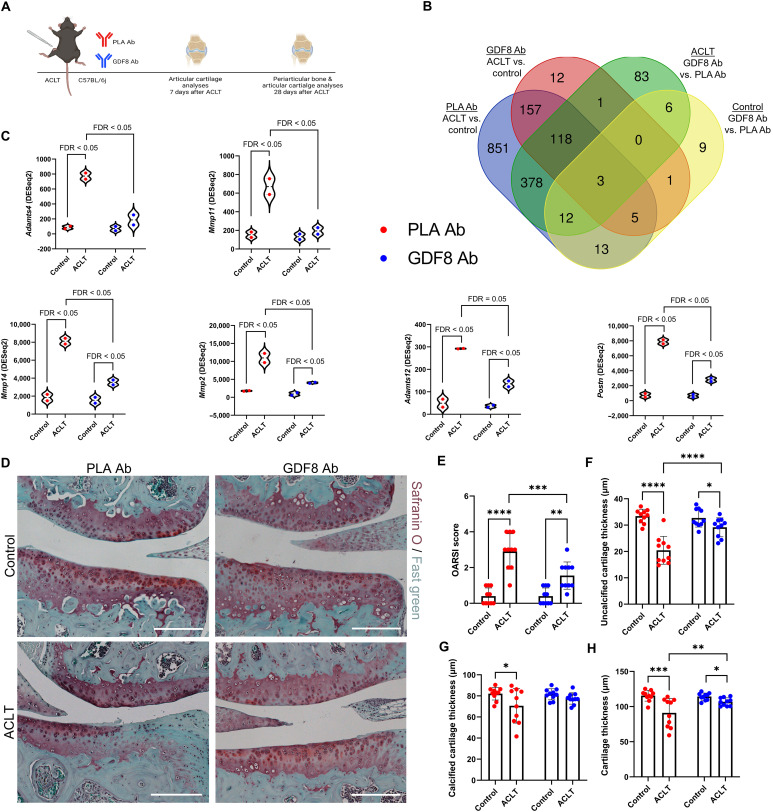
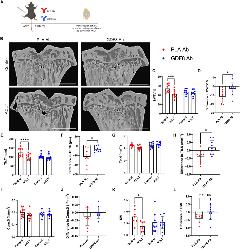
Musculoskeletal disorders contribute substantially to worldwide disability. Anterior cruciate ligament (ACL) tears result in unresolved muscle weakness and posttraumatic osteoarthritis (PTOA). Growth differentiation factor 8 (GDF8) has been implicated in the pathogenesis of musculoskeletal degeneration following ACL injury. We investigated GDF8 levels in ACL-injured human skeletal muscle and serum and tested a humanized monoclonal GDF8 antibody against a placebo in a mouse model of PTOA (surgically induced ACL tear). In patients, muscle GDF8 was predictive of atrophy, weakness, and periarticular bone loss 6 months following surgical ACL reconstruction. In mice, GDF8 antibody administration substantially mitigated muscle atrophy, weakness, and fibrosis. GDF8 antibody treatment rescued the skeletal muscle and articular cartilage transcriptomic response to ACL injury and attenuated PTOA severity and deficits in periarticular bone microarchitecture. Furthermore, GDF8 genetic deletion neutralized musculoskeletal deficits in response to ACL injury. Our findings support an opportunity for rapid targeting of GDF8 to enhance functional musculoskeletal recovery and mitigate the severity of PTOA after injury.


Figures

Fig. 1.
Fig. 1.. GDF8 is a molecular transducer of musculoskeletal deficits that are obstinate to surgical reconstruction and rehabilitation following ligamentous injury.
(A) Participants with ACL injury underwent measures of knee extensor dynamometry, quadriceps biopsies, and periarticular bone mineral density at indicated time points pre- and post-reconstruction. (B) Knee extension peak isometric torque and (C) rate of torque development are reduced after ACL injury and do not recover. (D) Representative immunohistochemistry (IHC) image of muscle fiber cross-sectional area (CSA). Scale bars, 200 μm. (E) Quadriceps muscle fiber atrophy occurs progressively after ACL injury and does not recover. (F) Representative image from dual-energy x-ray absorptiometry (DXA). The red box indicates distal femoral metaphysis; the yellow box indicates proximal tibial metaphysis. Scale bar, 4 cm. Bone mineral density is decreased in the (G) proximal tibial metaphysis and (H) distal femoral metaphysis 6 months following ACLR. (I) GDF8/myostatin expression is elevated in the quadriceps muscle after ACL injury and remains elevated after ACL surgical reconstruction (ACLR). (J) Greater loss of quadriceps/knee extension strength, quadriceps size, and periarticular bone density at 4 to 6 months after reconstruction are correlated with quadriceps muscle GDF8 expression after ACL injury. Scatterplots can be seen in fig. S3. N = 23 (B to E and I), 21 (G and H). **P < 0.01, ***P < 0.005, and ****P < 0.001 versus healthy via mixed effects model and Dunnett’s correction for multiple comparisons. AU, arbitrary units; GDF8KO, GDF8 knockout; BMD, bone mineral density; RTD, rate of torque development.
Fig. 2.
Fig. 2.. Humanized monoclonal antibody targeting GDF8 mitigates quadriceps atrophy and weakness and improves muscle quality following ACL injury.
(A) Study diagram; mice were treated biweekly. (B) PLA Ab–treated mice show quadriceps atrophy 1-week after ACL transection (ACLT) (drug × injury interaction, P = 0.002). (C) GDF8 Ab treatment rescues quadriceps fiber atrophy at 2 weeks after ACLT (drug × injury interaction, P = 0.024). (D) Representative tetanic graph from quadriceps peak isometric torque. (E) PLA Ab– and GDF8 Ab–treated mice show similar knee extension torque at 1 week after ACLT. (F) Knee extension weakness is mitigated by GDF8 Ab treatment 2 weeks after ACLT. Dashed lines in (E) and (F) represent mean values from uninjured control mice from historical laboratory data. (G) PLA Ab– and GDF8 Ab–treated mice show similar knee extension rates of torque development at 1 week after ACLT. (H) The knee extension rate of torque development is enhanced by GDF8 Ab treatment 2 weeks after ACLT injury. Dashed lines in (G) and (H) represent mean values from uninjured control mice from historical laboratory data. (I) Representative IHC images of quadriceps collagens 1 and 4 and collagen 1 (Col1)–GFP+ cells. scale bars, 100 μm. (J and K) GDF8 Ab treatment attenuates elevated abundance of Col1-GFP+ cells in quadriceps (J) 1 and (K) 2 weeks after ACLT [drug × injury interaction, P < 0.001 (J) and P = 0.019 (K)]. (L and M) GDF8 Ab treatment blocks the increase in collagens 1 and 4 in quadriceps (L) 1 and (M) 2 weeks after ACLT [drug × injury interaction, P = 0.006 (L) and P = 0.036 (M)]. N = 7 to 8 mice per group. *P < 0.05, **P < 0.01, ***P < 0.005, and ****P < 0.001 represent Šidák’s multiple comparison post hoc tests performed when significant interactions were detected via mixed models (B, C, J, K, L, and M) or independent sample t test (E to H). PLA Ab, placebo antibody; GDF8 Ab, GDF8 antibody; GFP, green fluorescent protein.
Fig. 3.
Fig. 3.. Treatment with anti-GDF8 antibody reduces muscle fibrogenic cell expansion and fibrosis following ACL injury.
(A and B) Representative images demonstrating Col1-GFP+ cells on (A) a muscle fiber and (B) following FACS isolation. Scale bars, 50 μm. (C) Representative FACS plot indicating muscle Col1-GFP+ cells; small inset indicates GFP+ cell absence in wild-type mice. (D) ACLT increases quadriceps Col1-GFP+ cell abundance in PLA Ab–treated mice (drug × injury interaction, P = 0.029). (E) Col1-GFP+ cells were gated by periostin expression level. (F and G) Representative histograms demonstrating periostin cell counts and GFP fluorescence intensity in ACLT limbs of PLA Ab– and GDF8 Ab–treated mice. (H and I) GDF8 Ab treatment lowers (H) raw and (I) relative cell abundance of periostin+; Col1-GFP+ cells in the ACLT quadriceps [drug × cell type interaction, P = 0.012 for both (H) and (I)]. (J and K) Representative images of isolated Col1-GFP+ cells stained for periostin. Scale bars, 100 μm. (L) GDF8 Ab treatment attenuates elevated periostin intensity in Col1-GFP+ cells 1 week after ACLT (drug × injury interaction, P = 0.011). (M and N) Representative IHC images denoting periostin, Col1-GFP, glycosaminoglycans (GAG), and 4′,6-diamidino-2-phenylindole (DAPI) in ACLT-injured quadriceps. White arrow: periostin+; Col1-GFP+ cell; yellow arrows: periostin; Col1-GFP+ cells. Scale bars, 50 μm. (O) GDF8 Ab treatment attenuates elevated frequency of quadriceps periostin+; Col1-GFP+ cells 1 week after ACLT (drug × injury interaction, P < 0.001). (P and Q) GDF8 Ab treatment rescues elevated (P) hydroxyproline abundance and (Q) pyridinoline (PYD) cross-linking in quadriceps muscle post-ACLT [drug × injury interaction, P = 0.002 for both (P) and (Q)]. N = 2 mice per group (E to I), 5 mice per group (C, D, and J to L), and 7 to 8 mice per group (M to Q). *P < 0.05, **P < 0.01, ***P < 0.005, and ****P < 0.001 represent Šidák’s multiple comparison post hoc tests performed when significant interactions were detected via mixed models. POSTN, periostin.
Fig. 4.
Fig. 4.. Transcriptomic analysis of most-changed genes by GDF8 inhibition in quadriceps muscle following ACL injury.
RNA-seq analysis was performed on quadriceps muscles from the study depicted in Fig. 2A at 1 week after ACLT (n = 5 mice per group; samples were not pooled). Analysis was performed on PLA Ab– and anti-GDF8 Ab–treated control and ACLT quadriceps. (A) Venn diagram depicting a number of genes differentially expressed by limb and treatment group. (B and C) Most activated (blue) and inhibited (red) pathways associated with GDF8 inhibition in the (B) ACLT quadriceps and (C) control quadriceps using REACTOME Pathway Knowledgebase. Pathways having false discovery rate (FDR) P < 0.05 for either direction are bright colors; pathways having FDR P > 0.05 for either direction are muted colors. (D) Expression of periostin (Postn) is elevated following ACLT and is mitigated by treatment with GDF8 Ab. (E) Expression of lysyl oxidase (Lox) is elevated following ACLT and is mitigated by treatment with GDF8 Ab. (F) Expression of Homer 2 (Homer2) is depressed following ACLT and is greater with anti-GDF8 Ab treatment. (G) Expression of nuclear receptor 4A3 (Nr4a3) is elevated in control and ACLT quadriceps following anti-GDF8 Ab treatment. Gene expression levels were calculated using the DESeq2 method. FDR < 0.05 denote specific comparisons.
Fig. 5.
Fig. 5.. GDF8 knockout preserves quadriceps size, strength, and quality following ACL injury.
(A) Study diagram; mice were studied at 7, 14, and 28 days after ACLT. (B to D) GDF8KO preserves quadriceps muscle fiber size (B) 7, (C) 14, and (D) 28 days after ACLT. (E) Representative tetanic graph from GDF8KO mouse knee extension peak isometric torque 14 days after ACLT. (F) Knee extension peak isometric torque is impaired 7 days after ACLT in GDF8KO mice (the dashed line represents the mean value from wild-type mice), but the strength impairment is rescued in GDF8KO mice 14 and 28 days after injury. (G) GDF8KO mitigates induction of pSMAD3 in the quadriceps 7 days following ACLT. (H) SMAD3 protein expression in GDF8KO mouse quadriceps muscle 7 days following ACLT. (I) pSMAD3 normalized to total SMAD3 in GDF8KO mouse quadriceps muscle 7 days following ACLT. (J) Representative IHC images of collagens 1 and 4 in control and ACLT quadriceps muscle. Scale bars, 100 μm. (K to M) GDF8KO protects against elevated collagen content in quadriceps (K) 7, (L) 14, and (M) 28 days after ACLT. N = 8 to 10 mice per group. *P < 0.05 via one-factor analysis of variance (ANOVA).
Fig. 6.
Fig. 6.. Treatment with anti-GDF8 antibody mitigates the severity of PTOA.
(A) Mice were treated biweekly; articular cartilage was collected from control and ACL transected knees at 1 week after ACLT for transcriptomic analysis and 4 weeks after ACLT for histopathology. (B) Eight mice were studied per group, and RNA was pooled from four mice to yield two biological replicates per group. A number of genes differentially expressed by limb (control and ACLT) and treatment (placebo- and GDF8-treated) groups were shown. (C) Expression of matrix metalloproteinases and factors affecting cartilage remodeling were elevated in articular cartilage 7 days after ACLT that were mitigated by GDF8 Ab treatment. Gene expression levels were calculated using DESeq2. (D) Representative images of safranin O and fast green from knee joints 4 weeks after ACLT. Scale bars, 200 μm. (E) Medial tibial plateau joint score (OARSI scoring system) shows the attenuation of PTOA severity in animals treated with anti-GDF8 Ab (drug × injury interaction, P = 0.005). (F) Uncalcified cartilage zones show thinning 28 days after ACLT that is protected by treatment with anti-GDF8 Ab (drug × injury interaction, P < 0.001). (G) Calcified cartilage zones show thinning 28 days after ACLT in placebo-treated animals (drug × injury interaction, P = 0.048). (H) Anti-GDF8 Ab treatment partially protects against total articular cartilage thinning 28 days after ACLT (drug × injury interaction, P = 0.019). N = 8 to 10 mice per group. *P < 0.05, **P < 0.01, ***P < 0.005, and ****P < 0.001 represent Šidák’s multiple comparison post hoc tests performed when significant interactions were detected via mixed models.
Fig. 7.
Fig. 7.. GDF8 antibody preserves periarticular bone microarchitecture following ACL injury.
(A) Study diagram; mice were treated biweekly with subcutaneous injections; whole knees were fixed for micro–computed tomography (micro-CT) analysis 4 weeks after ACLT. (B) Representative micro-CT images showing three-dimensional reconstruction of coronal sections. Scale bars, 1 mm. (C to L) Morphometric parameters of tibia subchondral trabecular bone. Anti-GDF8 treatment preserved bone volume fraction [BV/TV % (C and D), drug × injury interaction, P = 0.023], trabecular thickness [Tb.Th (E and F), drug × injury interaction, P = 0.017], and partially preserved trabecular number [Tb.N (G and H), drug × injury interaction, P = 0.020] 28 days after ACLT. There were no significant effects of ACLT or anti-GDF8 treatment on connectivity density [Conn.D (I and J)], but anti-GDF8 treatment protected against a loss of structure model index [SMI (K and L), drug × injury interaction, P = 0.047]. N = 9 mice per group. *P < 0.05, ***P < 0.005, and ****P < 0.001 represent Šidák’s multiple comparison post hoc tests performed when significant interactions were detected via mixed models (C, E, G, I, and K); *P < 0.05 via independent sample t test (D, F, H, J, and L).

Similar articles

Cited by

References

    1. C. J. Murray, C. Atkinson, K. Bhalla, G. Birbeck, R. Burstein, D. Chou, R. Dellavalle, G. Danaei, M. Ezzati, A. Fahimi, D. Flaxman, S. Foreman, E. Gabriel, N. Gakidou, S. Kassebaum, S. Khatibzadeh, S. Lim, E. Lipshultz, S. London, M. Lopez, F. MacIntyre, A. H. Mokdad, A. Moran, A. E. Moran, D. Mozaffarian, T. Murphy, M. Naghavi, C. Pope, T. Roberts, J. Salomon, D. C. Schwebel, S. Shahraz, D. A. Sleet, J. Murray, M. Abraham, K. Ali, D. H. Bartels, H. Chen, M. H. Criqui, J. Dahodwala, E. L. Ding, E. R. Dorsey, B. E. Ebel, S. Fahami, A. Flaxman, D. Flaxman, D. Gonzalez-Medina, B. Grant, H. Hagan, H. Hoffman, J. L. Leasher, J. Lin, R. Lozano, Y. Lu, L. Mallinger, M. M. McDermott, R. Micha, T. R. Miller, A. A. Mokdad, K. M. Narayan, S. B. Omer, P. M. Pelizzari, D. Phillips, D. Ranganathan, F. P. Rivara, U. Sampson, E. Sanman, A. Sapkota, S. Sharaz, R. Shivakoti, G. M. Singh, D. Singh, M. Tavakkoli, J. A. Towbin, J. D. Wilkinson, A. Zabetian, M. Alvardo, L. M. Baddour, E. J. Benjamin, I. Bolliger, E. Carnahan, S. S. Chugh, A. Cohen, K. E. Colson, L. T. Cooper, W. Couser, K. C. Dabhadkar, R. P. Dellavalle, D. Dicker, H. Duber, R. E. Engell, D. T. Felson, M. M. Finucane, T. Fleming, M. H. Forouzanfar, G. Freedman, M. K. Freeman, R. F. Gillum, R. Gosselin, H. R. Gutierrez, R. Havmoeller, K. H. Jacobsen, S. L. James, R. Jasrasaria, S. Jayarman, N. Johns, Q. Lan, M. Meltzer, G. A. Mensah, C. Michaud, C. Mock, T. E. Moffitt, R. G. Nelson, C. Olives, K. Ortblad, B. Ostro, M. Raju, H. Razavi, B. Ritz, R. L. Sacco, K. Shibuya, D. Silberberg, J. A. Singh, K. Steenland, J. A. Taylor, G. D. Thurston, M. S. Vavilala, T. Vos, G. R. Wagner, M. A. Weinstock, M. G. Weisskopf, S. Wulf; U.S. Burden of Disease Collaborators , The state of US health, 1990-2010: Burden of diseases, injuries, and risk factors. JAMA 310, 591–606 (2013). - PMC - PubMed
    1. I. J. Wallace, S. Worthington, D. T. Felson, R. D. Jurmain, K. T. Wren, H. Maijanen, R. J. Woods, D. E. Lieberman, Knee osteoarthritis has doubled in prevalence since the mid-20th century. Proc. Natl. Acad. Sci. U.S.A. 114, 9332–9336 (2017). - PMC - PubMed
    1. H. Kotlarz, C. L. Gunnarsson, H. Fang, J. A. Rizzo, Insurer and out-of-pocket costs of osteoarthritis in the US: Evidence from national survey data. Arthritis Rheum. 60, 3546–3553 (2009). - PubMed
    1. T. D. Brown, R. C. Johnston, C. L. Saltzman, J. L. Marsh, J. A. Buckwalter, Posttraumatic osteoarthritis: A first estimate of incidence, prevalence, and burden of disease. J. Orthop. Trauma 20, 739–744 (2006). - PubMed
    1. L. Y. Griffin, M. J. Albohm, E. A. Arendt, R. Bahr, B. D. Beynnon, M. Demaio, R. W. Dick, L. Engebretsen, W. E. Garrett Jr., J. A. Hannafin, T. E. Hewett, L. J. Huston, M. L. Ireland, R. J. Johnson, S. Lephart, B. R. Mandelbaum, B. J. Mann, P. H. Marks, S. W. Marshall, G. Myklebust, F. R. Noyes, C. Powers, C. Shields Jr., S. J. Shultz, H. Silvers, J. Slauterbeck, D. C. Taylor, C. C. Teitz, E. M. Wojtys, B. Yu, Understanding and preventing noncontact anterior cruciate ligament Injuries. Am. J. Sports Med. 34, 1512–1532 (2006). - PubMed

MeSH terms