. 2023 Dec;9(48):eadh5313.
doi: 10.1126/sciadv.adh5313. Epub 2023 Nov 29.

An injury-responsive mmp14b enhancer is required for heart regeneration

Affiliations

An injury-responsive mmp14b enhancer is required for heart regeneration

Ivana Zlatanova et al. Sci Adv. 2023 Dec.

Abstract

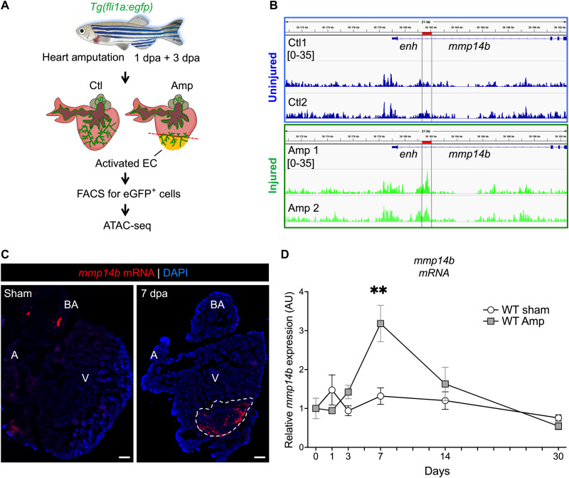
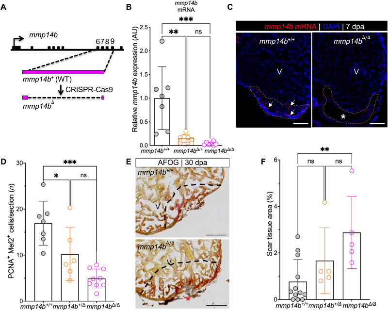
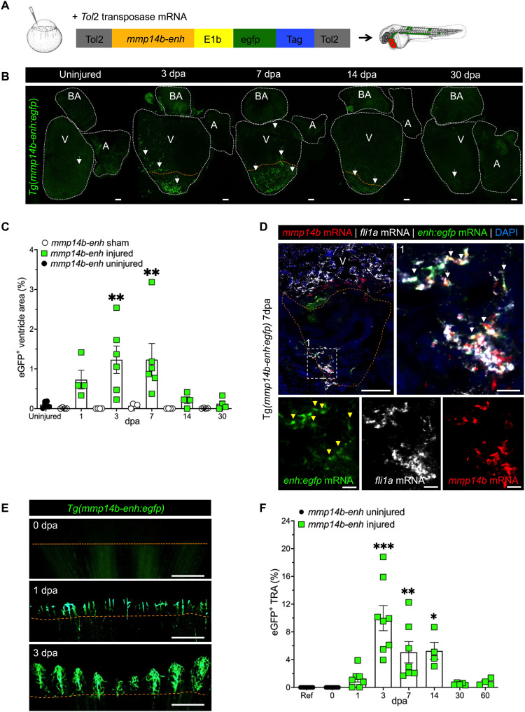
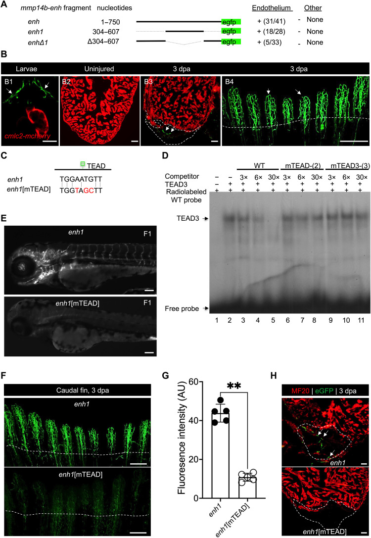
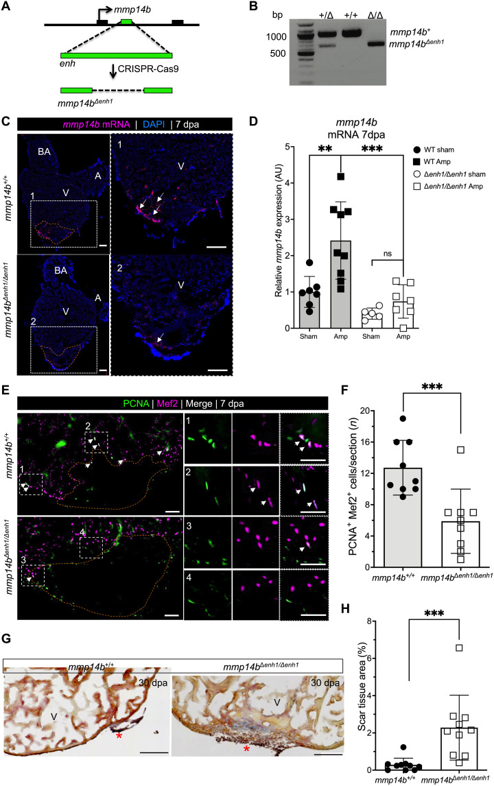
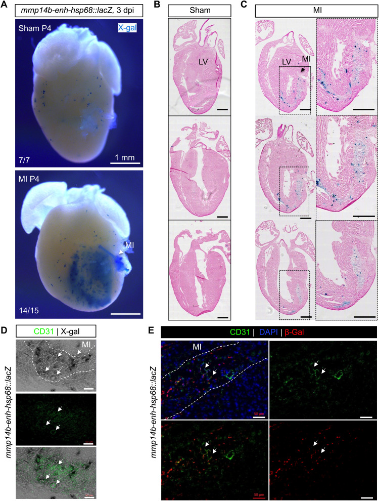
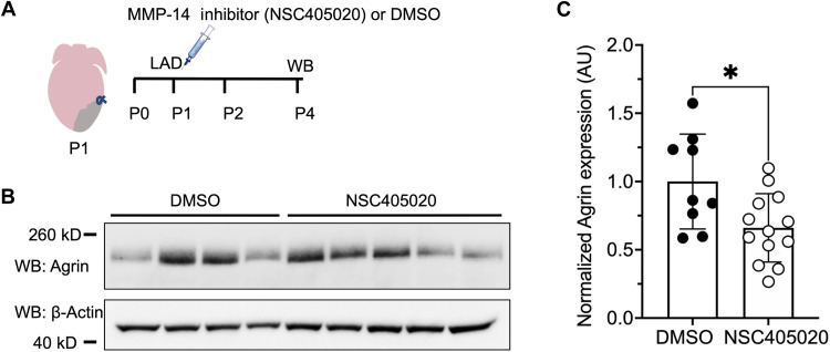
Mammals have limited capacity for heart regeneration, whereas zebrafish have extraordinary regeneration abilities. During zebrafish heart regeneration, endothelial cells promote cardiomyocyte cell cycle reentry and myocardial repair, but the mechanisms responsible for promoting an injury microenvironment conducive to regeneration remain incompletely defined. Here, we identify the matrix metalloproteinase Mmp14b as an essential regulator of heart regeneration. We identify a TEAD-dependent mmp14b endothelial enhancer induced by heart injury in zebrafish and mice, and we show that the enhancer is required for regeneration, supporting a role for Hippo signaling upstream of mmp14b. Last, we show that MMP-14 function in mice is important for the accumulation of Agrin, an essential regulator of neonatal mouse heart regeneration. These findings reveal mechanisms for extracellular matrix remodeling that promote heart regeneration.


Figures

Fig. 1.
Fig. 1.. mmp14b expression is induced in endothelial cells in response to heart injury in zebrafish.
(A) Experimental design for heart injury and subsequent ATAC-seq from cardiac ventricle endothelial cells. (B) Genome browser view showing ATAC-seq peak (enh) at 3 dpa in the mmp14b gene in uninjured (Ctl) and injured (Amp) eGFP+ cells. (C) RNAscope in situ hybridization for mmp14b expression in wild-type adult heart sections from uninjured (sham) and injured (7 dpa) hearts. mmp14b expression (red fluorescence) is evident in the injured area (dashed line). (D) Relative expression of mmp14b in wild-type zebrafish hearts expressed in arbitrary units (AU) without injury (sham) in white and injured (Amp) in gray at 1, 3, 7, 14, and 30 dpa as measured by qPCR. DAPI, 4′,6-diamidino-2-phenylindole; A, atrium; V, ventricle; BA, bulbous arteriosus. **P < 0.01.
Fig. 2.
Fig. 2.. Inactivation of the mmp14b gene results in reduced cardiomyocyte proliferation and impaired scar resolution in response to heart injury in zebrafish.
(A) Strategy used to create an mmp14b-null allele. (B) Relative mmp14b expression in mmp14b+/+, mmp14b+/Δ, and mmp14bΔ/Δ larvae as measured by qPCR. (C) RNAscope in situ hybridization for mmp14b mRNA in mmp14b+/+ and mmp14bΔ/Δ hearts at 7 dpa. Arrows, mmp14b expression in the wild-type mmp14b+/+ heart following injury. The asterisk denotes lack of evident mmp14b expression in the mmp14bΔ/Δ heart. (D) Cardiomyocyte proliferation indices at 7 dpa. (E) Representative frontal sections of mmp14b+/+ and mmp14bΔ/Δ hearts collected 30 dpa and stained with Acid Fushin Orange G (AFOG) to detect muscle (brown), fibrin (red), and collagen (blue). The asterisk highlights collagen-rich scar tissue. (F) Percent of ventricle composed of scar tissue at 30 dpa in mmp14b+/+, mmp14b+/Δ, and mmp14bΔ/Δ hearts. Data were collected for five to eight sections per heart and averaged to generate each data point. The black dashed lines mark the injured area. Scale bars, 100 μm. Statistical significance was determined using one-way analysis of variance (ANOVA) with Tukey’s multiple comparisons test. ns, not significant; *P < 0.05; **P < 0.01; ***P < 0.001.
Fig. 3.
Fig. 3.. Identification of an injury-responsive mmp14b endothelial enhancer.
(A) Schematic depicting generation of Tg(mmp14b-enh:egfp) zebrafish. (B) Representative whole-mount views of dissected Tg(mmp14b-enh:egfp) zebrafish hearts showing eGFP fluorescence in the vasculature at 3, 7, 14, and 30 dpa, compared to the background fluorescence in the uninjured heart (n = 9). White dashed lines outline the heart. Arrows highlight eGFP fluorescence in coronary endothelial cells. (C) Quantification of eGFP+ cells in Tg(mmp14b-enh:egfp) zebrafish in uninjured (black circles), sham-operated (open circles), and amputated (green squares) zebrafish ventricles at 1 to 30 dpa. (D) RNAscope in situ hybridization on frontal sections of Tg(mmp14b-enh:egfp) hearts at 7 dpa for mmp14b (red), egfp (green), and the endothelial marker fli1a (orange) (white arrowheads). Box 1 (white dashed line) represents the enlarged area to the right and in lower panels, which show individual color channels. (E) Fluorescent images of the caudal fin of Tg(mmp14b-enh:egfp) zebrafish at 0, 1, and 3 dpa show vascular-specific eGFP expression in newly formed vessels proximal to the amputation plane. (F) Vessel density in the total regenerated area (TRA) following caudal fin amputation from 0 to 60 days in Tg(mmp14b-enh:egfp) zebrafish (green squares). Uninjured Tg(mmp14b-enh:egfp) zebrafish (black circles) are shown as a reference. Orange dashed line approximates the amputation plane. Scale bars, 100 μm [(B) and (D)], 20 μm (D1), and 400 μm (E). Statistical significance was determined using one-way ANOVA with Tukey’s multiple comparisons test (C) or Dunnett’s test (F). *P < 0.05; **P < 0.01; ***P < 0.001.
Fig. 4.
Fig. 4.. Identification of a TEAD motif–dependent core region of mmp14b-enh.
(A) mmp14b-enh-egfp deletion constructs and their activity in transgenic zebrafish larvae at 72 hpf. Activity in endothelial cells and nonendothelial cells (other) is summarized. Numbers represent the fraction of larvae that exhibited activity in the vasculature. (B) Representative fluorescent images of Tg(mmp14b-enh1:egfp) transgenic zebrafish (B1), uninjured (B2), injured adult hearts (B3), and injured adult caudal fin (B4). eGFP fluorescence (green) indicates activity of the transgene. Cardiomyocytes in [(B1) to (B3)] are marked by cmlc2:mCherry (red). (C) TEAD site sequences in the mmp14b-enh1 and mmp14b-enh1[mTEAD] transgenes. (D) EMSA with recombinant TEAD3 and a radiolabeled, double-stranded mmp14b-enh1 TEAD site oligonucleotide probe. TEAD3 binding was efficiently competed by unlabeled, self-probe (lanes 3 to 5), but not versions in which two (lanes 6 to 8) or three (lanes 9 to 11) nucleotides were mutated. Unprogrammed reticulocyte lysate control (lane 1). WT, wild type. (E) Representative images of F1 generation Tg(mmp14b-enh1:egfp) and Tg(mmp14b-enh1:egfp[mTEAD]) zebrafish embryos at 72 hpf. (F) Representative images of caudal fins from stable transgenic lines, Tg(mmp14b-enh1:egfp) and Tg(mmp14b-enh1:egfp[mTEAD]) at 3 dpa. eGFP fluorescence (green) indicates activity of the transgenes. (G) Quantification of eGFP fluorescence intensity in AU in caudal fins 3 dpa from Tg(mmp14b-enh1:egfp) (closed circles) and Tg(mmp14b-enh1:egfp[mTEAD]) (open circles) zebrafish. (H) Heart sections from stable transgenic lines, Tg(mmp14b-enh1:egfp) and Tg(mmp14b-enh1:egfp[mTEAD]) at 3 dpa stained with anti-myosin (MF20, red) and anti-eGFP (green) antibodies. Dashed lines mark the amputation planes. White arrows denote endothelial expression. Scale bars, 50 μm [(B1), (B2), (B3), and (H)], 100 μm [(B4) and (E)], and 400 μm (F). Statistical significance in (G) was determined using Student’s t test. **P < 0.01.
Fig. 5.
Fig. 5.. mmp14benh1 is a bona fide mmp14b enhancer and is required for efficient heart regeneration.
(A) Strategy for deletion of the 304-bp mmp14b-enh1 sequence. (B) PCR genotyping of a mmp14b+/+, mmp14b+/Δenh1, and mmp14bΔenh1/Δenh1 zebrafish. (C) RNAscope in situ hybridization for endogenous mmp14b on frontal sections of injured mmp14b+/+ and mmp14bΔenh1/Δenh1 adult zebrafish hearts at 7 dpa. Boxed regions are enlarged in (1) and (2). Arrows highlight mmp14b expression (magenta). (D) Relative expression in AU of mmp14b in control (sham) and injured (Amp) mmp14b+/+ and mmp14bΔehn/1Δenh1 zebrafish hearts 7 dpa. (E) Sections of hearts from adult mmp14b+/+ and mmp14bΔenh1/Δenh1, subjected to ventricular amputation and immunostained for cardiomyocyte nuclei (⍺-Mef2; magenta) and cycling cells (⍺-PCNA; green). Boxed regions are enlarged in (1 to 4). Arrows highlight PCNA/Mef2 double-positive cardiomyocytes. (F) Cardiomyocyte proliferation indices at 7 dpa in mmp14b+/+ and mmp14bΔenh1/Δenh1 hearts. Proliferation data were collected for 10 sections per heart and averaged to generate each data point. (G) Frontal sections of adult mmp14b+/+ and mmp14bΔenh1/Δenh1 hearts 30 dpa stained with AFOG to detect muscle (brown), fibrin (red), and collagen (blue). The asterisk highlights collagen-rich scar tissue. (H) Residual scar as a percentage of total ventricular area at 30 dpa in mmp14b+/+ and mmp14bΔenh1/Δenh1 hearts; data were collected for four to six sections per heart and averaged to generate each data point. The orange dashed lines approximate the amputation area. Scale bars, 100 μm [(C) and (G)] and 50 μm (E). Statistical significance in (D) was determined using one-way ANOVA with Tukey’s multiple comparisons test. Statistical significance in (F) and (G) was determined using Student’s t test. **P < 0.01; ***P < 0.001.
Fig. 6.
Fig. 6.. Zebrafish mmp14b-enh is induced by myocardial injury in neonatal mice.
(A) Whole-mount and coronal sections (B and C) of representative mmp14b-enh-hsp68::lacZ hearts in control (sham) and injured myocardial infarction (MI) neonatal mice. Myocardial infarction was induced by permanent coronary ligation in neonatal mice on P1, and β-galactosidase (β-gal) expression was assessed by X-gal staining on P4. Sections were counterstained with hematoxylin and eosin. Note the activation of β-galactosidase (blue staining) in injured mmp14b-enh-hsp68::lacZ hearts (14 of 15) but not in sham-operated mmp14b-enh-hsp68::lacZ (7 of 7). Boxes in [(C), left] are shown at higher magnification in [(C), right]. LV, left ventricle. (D and E) Coronal sections of mmp14b-enh-hsp68::lacZ neonatal mouse hearts stained for X-gal (gray) (D) or immunostained for β-galactosidase (red) (E) and immunostained for the endothelial cell marker CD31 (green) at 3 dpi. Colocalization of X-gal and CD31 is shown in the bottom of (D). Colocalized expression is highlighted by white arrows. The infarcted area is outlined with a white dashed line. DAPI staining (blue) is shown in (E). Scale bars, 500 μm [(B) and (C)] and 50 μm [(D) and (E)].
Fig. 7.
Fig. 7.. MMP-14 activity facilitates Agrin availability in the extracellular matrix of neonatal mice.
(A) Schematic of the experimental design for neonatal mouse heart injury and MMP-14 inhibitor treatment. Myocardial injury by coronary artery ligation was conducted on P1, mice were simultaneously treated with a single intraperitoneal injection of 20 μM NSC405020 or DMSO (vehicle control), and hearts were collected on P4 for analysis by Western blot (WB). (B) Western blots for Agrin and β-actin from total heart tissue 3 dpi. (C) Quantification of the 260-kD isoform of Agrin protein normalized to β-actin and expressed in AU in neonatal mouse hearts following myocardial injury (3 dpi) and treatment with either DMSO or NSC405020. Statistical significance was determined using Student’s t test. *P < 0.05.

References

    1. E. J. Benjamin, S. S. Virani, C. W. Callaway, A. M. Chamberlain, A. R. Chang, S. Cheng, S. E. Chiuve, M. Cushman, F. N. Delling, R. Deo, S. D. de Ferranti, J. F. Ferguson, M. Fornage, C. Gillespie, C. R. Isasi, M. C. Jiménez, L. C. Jordan, S. E. Judd, D. Lackland, J. H. Lichtman, L. Lisabeth, S. Liu, C. T. Longenecker, P. L. Lutsey, J. S. Mackey, D. B. Matchar, K. Matsushita, M. E. Mussolino, K. Nasir, M. O'Flaherty, L. P. Palaniappan, A. Pandey, D. K. Pandey, M. J. Reeves, M. D. Ritchey, C. J. Rodriguez, G. A. Roth, W. D. Rosamond, U. K. A. Sampson, G. M. Satou, S. H. Shah, N. L. Spartano, D. L. Tirschwell, C. W. Tsao, J. H. Voeks, J. Z. Willey, J. T. Wilkins, J. H. Wu, H. M. Alger, S. S. Wong, P. Muntner, Heart Disease and Stroke Statistics-2018 Update: A report from the American Heart Association. Circulation 137, e67–e492 (2018). - PubMed
    1. J. Johnson, S. Mohsin, S. R. Houser, Cardiomyocyte proliferation as a source of new myocyte development in the adult heart. Int. J. Mol. Sci. 22, 7764 (2021). - PMC - PubMed
    1. W. Wang, C. K. Hu, A. Zeng, D. Alegre, D. Hu, K. Gotting, A. Ortega Granillo, Y. Wang, S. Robb, R. Schnittker, S. Zhang, D. Alegre, H. Li, E. Ross, N. Zhang, A. Brunet, A. Sánchez Alvarado, Changes in regeneration-responsive enhancers shape regenerative capacities in vertebrates. Science 369, eaaz3090 (2020). - PMC - PubMed
    1. T. Hoang, J. Wang, P. Boyd, F. Wang, C. Santiago, L. Jiang, S. Yoo, M. Lahne, L. J. Todd, M. Jia, C. Saez, C. Keuthan, I. Palazzo, N. Squires, W. A. Campbell, F. Rajaii, T. Parayil, V. Trinh, D. W. Kim, G. Wang, L. J. Campbell, J. Ash, A. J. Fischer, D. R. Hyde, J. Qian, S. Blackshaw, Gene regulatory networks controlling vertebrate retinal regeneration. Science 370, eabb8598 (2020). - PMC - PubMed
    1. T. Gerber, P. Murawala, D. Knapp, W. Masselink, M. Schuez, S. Hermann, M. Gac-Santel, S. Nowoshilow, J. Kageyama, S. Khattak, J. D. Currie, J. G. Camp, E. M. Tanaka, B. Treutlein, Single-cell analysis uncovers convergence of cell identities during axolotl limb regeneration. Science 362, eaaq0681 (2018). - PMC - PubMed

Substances