

Risk Factors for Bleeding in Coronavirus Disease 2019 Patients on Extracorporeal Membrane Oxygenation and Effects of Transcatheter Arterial Embolization for Hemostasis
- PMID: 38020462
- PMCID: PMC10681754
- DOI: 10.22575/interventionalradiology.2022-0043
Risk Factors for Bleeding in Coronavirus Disease 2019 Patients on Extracorporeal Membrane Oxygenation and Effects of Transcatheter Arterial Embolization for Hemostasis
Abstract
Purpose: To evaluate risk factors for bleeding events in coronavirus disease 2019 (COVID-19) patients on extracorporeal membrane oxygenation (ECMO) and to share the initial results of transcatheter arterial embolization (TAE) for hemostasis.
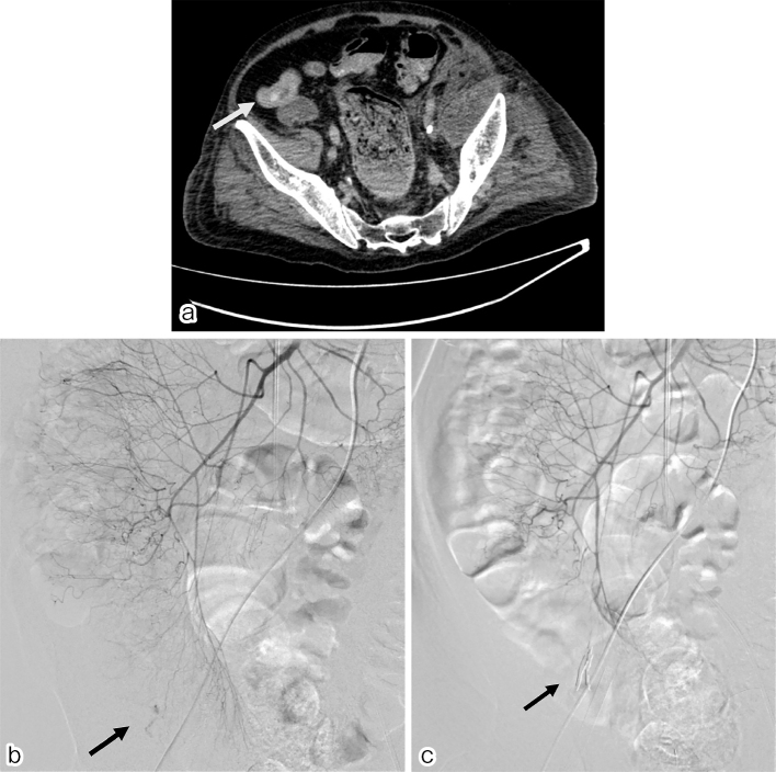
Material and methods: Forty-three COVID-19 patients who received ECMO from May 2020 to September 2021 were enrolled in this study. Patients with sudden onset anemia immediately underwent computed tomography to assess bleeding. We compared laboratory data, duration of ECMO, hospitalization period, and fatality of patients' groups with and without significant hemorrhagic events using the chi-square test and Mann-Whitney U test. We also assessed the results of TAE in patients who received hemostasis.
Results: A total of 25 bleeding events occurred in 24 of the 43 patients. Age was a risk factor for bleeding events and fatality. The average duration of ECMO and hospitalization period were significantly longer in those with bleeding events (42.9 and 54.3 days) than in those without bleeding events (16.2 and 25.0 days) (p < 0.05). In addition, those with bleeding had higher fatality (45.8%) than those without (15.8%) (p < 0.05). Active extravasation was confirmed for 5 events in 4 of 24 patients. TAE was attempted and performed successfully in all but one of these four cases, in whom bleeding ceased spontaneously.
Conclusions: Elderly COVID-19 patients on ECMO had a greater risk of bleeding complications and fatal outcomes. TAE was effective in providing prompt hemostasis for patients who have the treatment indication.
Keywords: bleeding; coronavirus disease 2019 (COVID-19); extracorporeal membrane oxygenation (ECMO); extravasation; transcatheter arterial embolization (TAE).
© 2023 Japanese Society of Interventional Radiology.
Conflict of interest statement
None
Figures


Similar articles
-
Efficacy of transcatheter arterial embolization for first-line treatment of colonic diverticular bleeding with extravasation on contrast-enhanced computed tomography.Medicine (Baltimore). 2022 Nov 4;101(44):e31442. doi: 10.1097/MD.0000000000031442. Medicine (Baltimore). 2022. PMID: 36343028 Free PMC article.
-
Video-assisted thoracic surgery in critically ill COVID-19 patients on venovenous extracorporeal membrane oxygenation.Perfusion. 2023 Nov;38(8):1577-1583. doi: 10.1177/02676591221119319. Epub 2022 Aug 15. Perfusion. 2023. PMID: 35969115 Free PMC article.
-
The efficacy of transcatheter arterial embolization as the first-choice treatment after failure of endoscopic hemostasis and endoscopic treatment resistance factors.Dig Endosc. 2012 Sep;24(5):364-9. doi: 10.1111/j.1443-1661.2012.01285.x. Epub 2012 Apr 2. Dig Endosc. 2012. PMID: 22925291
-
Transcatheter arterial embolization versus surgery for refractory non-variceal upper gastrointestinal bleeding: a meta-analysis.World J Emerg Surg. 2019 Feb 1;14:3. doi: 10.1186/s13017-019-0223-8. eCollection 2019. World J Emerg Surg. 2019. PMID: 30733822 Free PMC article.
-
In-hospital outcomes after emergency or prophylactic veno-arterial extracorporeal membrane oxygenation during transcatheter aortic valve implantation: a comprehensive review of the literature.Perfusion. 2019 Jul;34(5):354-363. doi: 10.1177/0267659118816555. Epub 2019 Jan 11. Perfusion. 2019. PMID: 30632894 Review.
Cited by
-
Transarterial embolization to treat a massive hemothorax during mechanical circulatory support via puncturing of the extracorporeal membrane oxygenation circuit.CVIR Endovasc. 2024 May 21;7(1):48. doi: 10.1186/s42155-024-00460-8. CVIR Endovasc. 2024. PMID: 38769160 Free PMC article.
References
-
- Hadaya J, Benharash P. Extracorporeal membrane oxygenation. JAMA. 2020; 323: 2536. - PubMed