. 2023 Oct 16;3(11):100420.
doi: 10.1016/j.xgen.2023.100420. eCollection 2023 Nov 8.

Identification of a regulatory pathway governing TRAF1 via an arthritis-associated non-coding variant

Affiliations

Identification of a regulatory pathway governing TRAF1 via an arthritis-associated non-coding variant

Qiang Wang et al. Cell Genom. .

Erratum in

  • Identification of a regulatory pathway governing TRAF1 via an arthritis-associated non-coding variant.
    Wang Q, Martínez-Bonet M, Kim T, Sparks JA, Ishigaki K, Chen X, Sudman M, Aguiar V, Sim S, Hernandez MC, Chiu DJ, Wactor A, Wauford B, Marion MC, Gutierrez-Arcelus M, Bowes J, Eyre S, Nordal E, Prahalad S, Rygg M, Videm V, Raychaudhuri S, Weirauch MT, Langefeld CD, Thompson SD, Nigrovic PA. Wang Q, et al. Cell Genom. 2024 Feb 14;4(2):100502. doi: 10.1016/j.xgen.2024.100502. Cell Genom. 2024. PMID: 38359789 Free PMC article. No abstract available.

Abstract

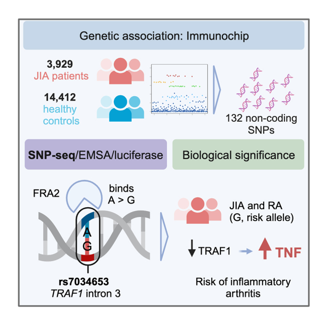
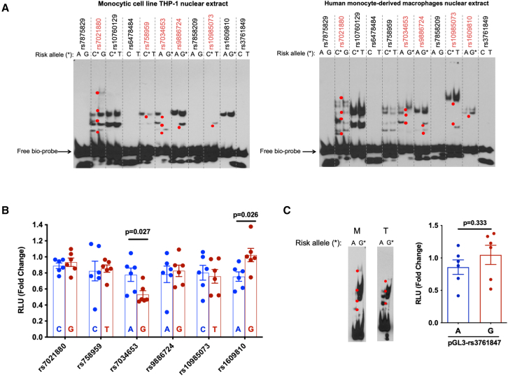
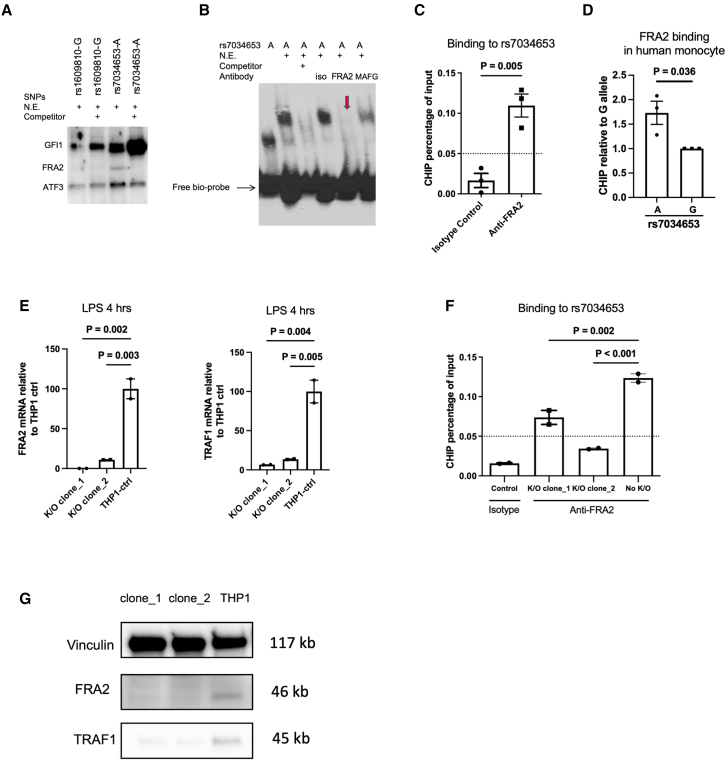
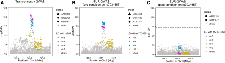
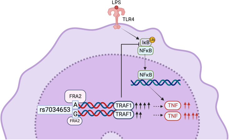
TRAF1/C5 was among the first loci shown to confer risk for inflammatory arthritis in the absence of an associated coding variant, but its genetic mechanism remains undefined. Using Immunochip data from 3,939 patients with juvenile idiopathic arthritis (JIA) and 14,412 control individuals, we identified 132 plausible common non-coding variants, reduced serially by single-nucleotide polymorphism sequencing (SNP-seq), electrophoretic mobility shift, and luciferase studies to the single variant rs7034653 in the third intron of TRAF1. Genetically manipulated experimental cells and primary monocytes from genotyped donors establish that the risk G allele reduces binding of Fos-related antigen 2 (FRA2), encoded by FOSL2, resulting in reduced TRAF1 expression and enhanced tumor necrosis factor (TNF) production. Conditioning on this JIA variant eliminated attributable risk for rheumatoid arthritis, implicating a mechanism shared across the arthritis spectrum. These findings reveal that rs7034653, FRA2, and TRAF1 mediate a pathway through which a non-coding functional variant drives risk of inflammatory arthritis in children and adults.

Keywords: C5; FRA2; TNF; TRAF1; genome-wide association study; juvenile idiopathic arthritis; monocyte; non-coding variant; rheumatoid arthritis.


Conflict of interest statement

The authors declare no competing interests.

Figures

None
Graphical abstract
Figure 1
Figure 1
Analysis of genetic association at the TRAF1/C5 locus in JIA To define the non-coding variant within TRAF1/C5, we employed Immunochip data from 3,939 patients with oligoarticular and seronegative polyarticular JIA and 14,412 control subjects. SNP rs7039505 exhibited confirmed significance, p = 2.39 × 10−7 via logistic regression, odds ratio = 1.15, 95% confidence interval 1.09–1.21, Benjamini-Hochberg false discovery rate pFDR = 9.33 × 10−6 adjusted for the number of SNPs that passed quality control, defined as high-quality clustering, call rates >95%, meeting Hardy-Weinberg equilibrium expectations, and no differential missingness between case subjects and control subjects.
Figure 2
Figure 2
Colocalization between JIA GWAS and eQTL signals We tested for colocalization of the JIA association with eQTLs for 11 genes in the TRAF1/C5 region. Results are shown for eQTL Catalog datasets (y axis) with a probability of colocalization from coloc (PP4, posterior probability of hypothesis 4, that a single variant explains both genetic and eQTL signals, x axis) greater than 0.5. eQTL datasets are ordered by their maximum PP4, and datasets for monocytes and macrophages are highlighted in red. Genes with no PP4 >0.5 are not shown (white points in legend).
Figure 3
Figure 3
Screening candidate regulatory variants within TRAF1/C5 by SNP-seq (A) SNP-seq. A 31 bp sequence centered upon the SNP is flanked by two type IIS restriction enzyme (IISRE) binding sites. A primer is included for high-throughput sequencing. SNPs that fail to bind transcription factors (TFs) or other regulatory proteins are negatively selected. Protected constructs are amplified using primers as per Table S2. Bio, biotin; NGS, next-generation sequencing. Created with BioRender.com. (B) Correlation analysis of NGS data for each replicate of SNP-seq, using non-linear regression. (C) Read count ratio between samples and controls at cycle 10 for 24 protected SNPs, with alleles 1 and 2 for each SNP shown in red and blue, respectively. 16 SNPs exhibited allele-specific protection. Statistical analysis was performed using unpaired t tests (n = 3 replicates, ∗p < 0.05). (D) Top four SNPs showing an increment in allele-specific protection across cycles 4, 6, 8, and 10. A linear model for the ratio of reads in sample vs. control for alleles 1 and 2, with an absolute slope greater than 0.05. (E) HaploReg annotations for the 11 candidate regulatory SNPs selected from SNP-seq.
Figure 4
Figure 4
Experimental assessment of candidate regulatory SNPs and impact on inflammatory cytokine production by primary monocytes (A) EMSA for 11 candidate regulatory SNPs. Allele-specific gel shift/binding is highlighted with red dots. Left: result with nuclear extract from the monocytic cell line THP-1; right: result with nuclear extract from human monocyte-derived macrophages (representative of n = 3 independent biological replicates with similar results). (B) Luciferase reporter assay showing relative fluorescence in human THP-1 cells between the non-risk (blue) and risk (red) alleles of 6 candidate regulatory SNPs from EMSA (mean ± SEM n = 6 biological replicates, t test with two tails without correction for multiple hypothesis testing); we proceeded with analysis of rs7034653. (C) EMSA and luciferase reporter assay for SNP rs3761847. M indicates human monocyte-derived macrophages, and T indicates THP-1 cells. For luciferase assays, histogram shows mean ± SEM, n = 6 biological replicates, t test with two tails without correction for multiple hypothesis testing. (D–F) Purified monocytes from healthy human donors were treated or not with LPS (100 ng/mL for the duration shown). (D) Histograms by flow cytometry showing the time course of induction of TRAF1 by LPS in purified monocytes from one representative donor (AG genotype at rs7034653). (E) Mean fluorescence intensity (MFI) of TRAF1 in purified monocytes of subjects with the AA, AG, and GG genotypes at rs7034653. (F) ELISA measurements of TNF in monocyte supernatants after LPS stimulation. Each symbol represents one donor; n = 13, 13, and 12 human subjects of genotypes AA, AG, and GG, respectively. Statistical analysis was performed using one-way ANOVA with multiple comparisons.
Figure 5
Figure 5
FRA2 binding to rs7034653 regulates TRAF1 expression (A) Oligonucleotide pull-down western blot assay showing that FRA2 binds specifically to rs7034653-A; binding is eliminated by 30-fold excess non-biotinylated rs7034653-A competitor. GFI1 and ATF3 bind to all samples with or without non-biotinylated competitors. (B) EMSA supershift showing specific binding of rs7034653-A to FRA2. The addition of antibody to FRA2 in the oligo/nuclear extract (THP-1) mixture caused the disappearance of the shift band (arrow), whereas isotype control antibody and antibody to irrelevant control TF MAFG did not. (C) ChIP-qPCR showing FRA2 binding to rs7034653 in THP-1 cells (mean ± SEM, n = 3). (D) Allele discrimination ChIP-qPCR showing that FRA2 preferentially binds to A allele over G allele of rs7034653 in unstimulated human monocytes heterozygous at rs7034653 (mean ± SEM, n = 3). (E and G) CRISPR-Cas9 FRA2 knockout THP-1 clones show decreased FRA2 and TRAF1 mRNA and protein expression compared with negative single guide (sg) RNA-treated THP-1 control (mean ± SEM, n = 2). (F) CRISPR-Cas9 FRA2 knockout THP-1 clones show decreased FRA2 binding to rs7034653 compared with negative sgRNA-treated THP-1 control (mean ± SEM, n = 2). (A), (B), and (E)–(G) are representative of duplicate biological replicates, and (C) and (D) are representative of triplicate biological replicates. Statistical analysis methods used for (C) and (D) were unpaired t test with two tails without correction and for (E) and (F) were one-way ANOVA with multiple comparisons.
Figure 6
Figure 6
Conditional analysis on rs7034653 confirms no residual risk in RA (A) TRAF1/C5 locuszoom plot in trans-ancestry GWAS of RA. (B) Pre-conditional analysis locuszoom plot in TRAF1/C5 in the European-ancestry cohort, comparable to the JIA Immunochip population. (A and B) p values for were calculated by a fixed effect meta-analysis of statistics from logistic regression tests. (C) Post-conditional analysis in the European population detected no residual RA risk. Conditional analysis using a logistic regression model was conducted in each cohort of the 25 European-ancestry GWAS, and the results were meta-analyzed using the inverse-variance weighted fixed-effect model.
Figure 7
Figure 7
Allelic variation at rs7034653 modulates TNF production through differential binding of FRA2 to regulate expression of TRAF1 FRA2 exhibits greater affinity for the protective A allele than the risk G allele of rs7034653, leading to enhanced production of the negative regulatory protein TRAF1 and lower production of the pro-inflammatory mediator TNF. Inhibition of NF-κB by TRAF1 occurs indirectly, mediated through interaction with the linear ubiquitin chain assembly complex (LUBAC) to block linear ubiquitination of IKKγ, preventing the degradation of the IKK complex required for entry of NF-κB into the nucleus. Created with BioRender.com.

References

    1. Farh K.K.H., Marson A., Zhu J., Kleinewietfeld M., Housley W.J., Beik S., Shoresh N., Whitton H., Ryan R.J.H., Shishkin A.A., et al. Genetic and epigenetic fine mapping of causal autoimmune disease variants. Nature. 2015;518:337–343. doi: 10.1038/nature13835. - DOI - PMC - PubMed
    1. Li G., Martínez-Bonet M., Wu D., Yang Y., Cui J., Nguyen H.N., Cunin P., Levescot A., Bai M., Westra H.J., et al. High-throughput identification of noncoding functional SNPs via type IIS enzyme restriction. Nat. Genet. 2018;50:1180–1188. doi: 10.1038/s41588-018-0159-z. - DOI - PMC - PubMed
    1. Lu X., Chen X., Forney C., Donmez O., Miller D., Parameswaran S., Hong T., Huang Y., Pujato M., Cazares T., et al. Global discovery of lupus genetic risk variant allelic enhancer activity. Nat. Commun. 2021;12:1611. doi: 10.1038/s41467-021-21854-5. - DOI - PMC - PubMed
    1. Moore J.H., Asselbergs F.W., Williams S.M. Bioinformatics challenges for genome-wide association studies. Bioinformatics. 2010;26:445–455. doi: 10.1093/bioinformatics/btp713. - DOI - PMC - PubMed
    1. Zhang Q., Bastard P., Liu Z., Le Pen J., Moncada-Velez M., Chen J., Ogishi M., Sabli I.K.D., Hodeib S., Korol C., et al. Inborn errors of type I IFN immunity in patients with life-threatening COVID-19. Science. 2020;370:eabd4570. doi: 10.1126/science.abd4570. - DOI - PMC - PubMed

LinkOut - more resources