

Case Reports
doi: 10.7759/cureus.47367.
eCollection 2023 Oct.
Unprovoked Spontaneous Kidney Rupture (Wunderlich's Syndrome) Managed by Renal Artery Embolization
Affiliations
- PMID: 38021993
- PMCID: PMC10657482
- DOI: 10.7759/cureus.47367
Item in Clipboard
Case Reports
Unprovoked Spontaneous Kidney Rupture (Wunderlich's Syndrome) Managed by Renal Artery Embolization
Cureus.
.
Abstract
Wunderlich's syndrome is a rare, unfamiliar disease that can present with flank pain, flank mass, and hypovolemic shock without any history of trauma. In this article, we present a sudden, unprovoked kidney rupture managed by renal artery embolization. This report emphasizes the importance of early referral and prompt management, which can be lifesaving.
Keywords: active bleeding; flank pain; renal artery embolization; spontaneous kidney rupture; wunderlich's syndrome.
Copyright © 2023, Alzahrani et al.
Conflict of interest statement
The authors have declared that no competing interests exist.
Figures

Seen is an unremarkable right kidney (A) apart from the minimal perinephric free fluid (white arrow in image B).

Seen is a shattered kidney with cortical distortion and active contrast extravasation (white arrows in images A, B, and C).

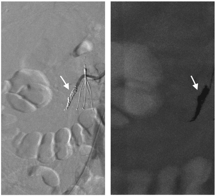

A and B: An infected hematoma with air pockets (white arrows), C: Coil embolization (arrowhead)
References
-
- Wunderlich syndrome: cross-sectional imaging review. Katabathina VS, Katre R, Prasad SR, Surabhi VR, Shanbhogue AK, Sunnapwar A. J Comput Assist Tomogr. 2011;35:425–433. - PubMed
-
- Sudden onset flank pain: spontaneous renal rupture. Grubb SM, Stuart JI, Harper HM. Am J Emerg Med. 2017;35:1787. - PubMed
-
- A rare case of spontaneous parenchymal kidney explosion in a patient with ureteral obstruction caused by a single stone. Chiancone F, Meccariello C, Ferraiuolo M, De Marco GP, Fedelini M, Langella NA, Fedelini P. Urologia. 2021;88:386–388. - PubMed
Publication types
LinkOut - more resources
Full Text Sources