

Prevalence and Clinical Significance of Incidental Findings in the Maxillofacial Complex of Adolescent Orthodontic Patients: A Retrospective Cone Beam Computed Tomography Analysis
- PMID: 38022275
- PMCID: PMC10663048
- DOI: 10.7759/cureus.47480
Prevalence and Clinical Significance of Incidental Findings in the Maxillofacial Complex of Adolescent Orthodontic Patients: A Retrospective Cone Beam Computed Tomography Analysis
Abstract
Objectives: The aim of this study was to determine the prevalence and severity of incidental findings in the maxillofacial complex of orthodontic patients imaged with cone beam computed tomography (CBCT) and assign those findings an appropriate clinical significance.
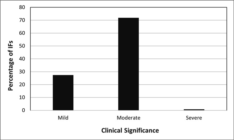
Methodology: Incidental findings (IF) were identified in 250 CBCT scans of adolescent orthodontic patients (aged 13-18 years) with a large field-of-view and categorized based on their anatomic location and placed into one of six subgroups based on anatomic region: i) sino-nasal, ii) dentoalveolar, iii) nasooropharyngeal airway, iv) temporomandibular joint, v) neck, vi) calcifications, and vi) miscellaneous findings. Additionally, findings were assigned a clinical significance score based on severity on a scale of mild, moderate and severe. Mild IF was defined as an IF that does not require any further investigation or referral. Moderate IF was defined as an IF that has the tendency to become clinically significant and should be observed periodically. IFs that warrant further investigation and/or intervention were designated as severe.
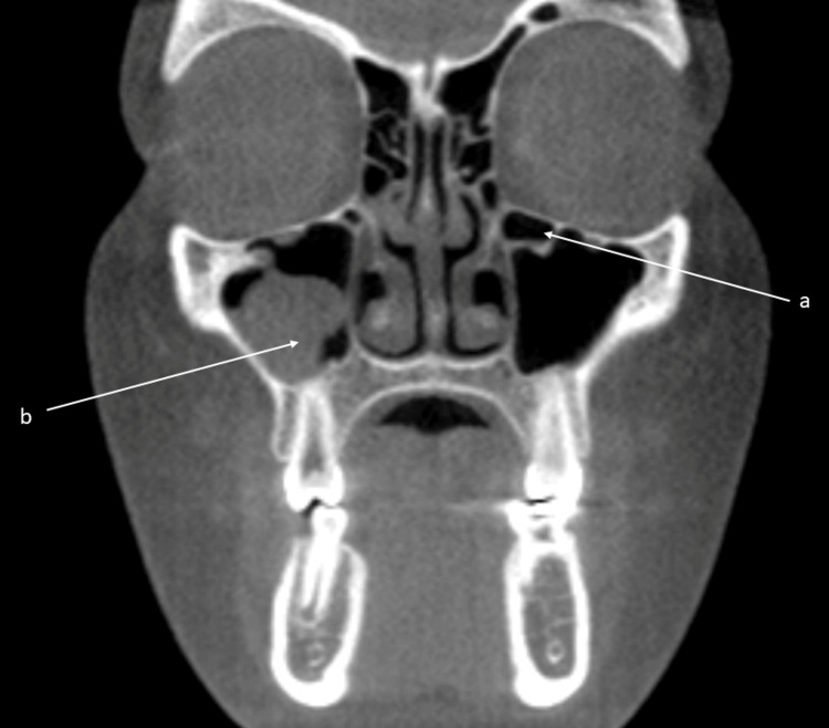
Results: The percentage of IFs in sino-nasal and dento-alveolar regions were 44.7% and 19.1% respectively. The percentage of IFs with mild, moderate, and severe clinical significance were 27%, 72%, and 1%, respectively. Out of the IFs involving calcifications, 80.8% were stylohyoid calcifications and <1% were cranial cavity IFs such as petroclinoid calcifications and falx cerebri calcifications. Among the sino-nasal findings, 1.2% were identified as severe.
Conclusion: The sino-nasal region had the highest frequency of IFs. Understanding the prevalence of incidental findings and its clinical relevance is important for clinicians to allow for appropriate monitoring and timely treatment of patients.
Keywords: 3d imaging; adolescent orthodontics; cone-beam computed tomography (cbct); incidental findings; orthodontics.
Copyright © 2023, Etemad et al.
Conflict of interest statement
The authors have declared that no competing interests exist.
Figures
Similar articles
-
Incidental findings on craniomaxillofacial cone beam computed tomography in orthodontic patients.Int J Comput Dent. 2019;22(2):149-162. Int J Comput Dent. 2019. PMID: 31134221
-
A systematic review on incidental findings in cone beam computed tomography (CBCT) scans.Dentomaxillofac Radiol. 2019 Oct;48(7):20180396. doi: 10.1259/dmfr.20180396. Epub 2019 Jun 28. Dentomaxillofac Radiol. 2019. PMID: 31216179 Free PMC article.
-
Study of the frequency and location of incidental findings of the maxillofacial region in different fields of view in CBCT scans.Dentomaxillofac Radiol. 2017 Jan;46(1):20160215. doi: 10.1259/dmfr.20160215. Epub 2016 Oct 5. Dentomaxillofac Radiol. 2017. PMID: 27604390 Free PMC article.
-
Retrospective Evaluation of Incidental Findings of Temporomandibular Joint Region in CBCT Scans.J Contemp Dent Pract. 2021 Dec 1;22(12):1393-1398. J Contemp Dent Pract. 2021. PMID: 35656676
-
Comparison of incidental findings on cone beam computed tomographic and 2-dimensional images.Gen Dent. 2023 Jul-Aug;71(4):64-71. Gen Dent. 2023. PMID: 37358586 Review.
Cited by
-
Incidental findings from cone-beam computed tomography in children and adolescents: a systematic review.Eur Arch Paediatr Dent. 2025 Jan 17. doi: 10.1007/s40368-025-00999-7. Online ahead of print. Eur Arch Paediatr Dent. 2025. PMID: 39820816
References
-
- What is cone-beam CT and how does it work? Scarfe WC, Farman AG. Dent Clin North Am. 2008;52:707–730. - PubMed
-
- Managing incidental findings on abdominal CT: white paper of the ACR incidental findings committee. Berland LL, Silverman SG, Gore RM, et al. J Am Coll Radiol. 2010;7:754–773. - PubMed
-
- Langland OE, Langlais RP. Baltimore: Lippincott Williams & Wilkins; 1997. Principles of Dental Imaging.
LinkOut - more resources
Full Text Sources
Miscellaneous