Review
. 2023 Oct 20;14(45):12815-12849.
doi: 10.1039/d3sc04052f. eCollection 2023 Nov 22.

Fluorescence-readout as a powerful macromolecular characterisation tool

Affiliations
Review

Fluorescence-readout as a powerful macromolecular characterisation tool

Xingyu Wu et al. Chem Sci. .

Abstract

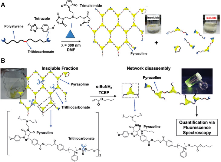
The last few decades have witnessed significant progress in synthetic macromolecular chemistry, which can provide access to diverse macromolecules with varying structural complexities, topology and functionalities, bringing us closer to the aim of controlling soft matter material properties with molecular precision. To reach this goal, the development of advanced analytical techniques, allowing for micro-, molecular level and real-time investigation, is essential. Due to their appealing features, including high sensitivity, large contrast, fast and real-time response, as well as non-invasive characteristics, fluorescence-based techniques have emerged as a powerful tool for macromolecular characterisation to provide detailed information and give new and deep insights beyond those offered by commonly applied analytical methods. Herein, we critically examine how fluorescence phenomena, principles and techniques can be effectively exploited to characterise macromolecules and soft matter materials and to further unravel their constitution, by highlighting representative examples of recent advances across major areas of polymer and materials science, ranging from polymer molecular weight and conversion, architecture, conformation to polymer self-assembly to surfaces, gels and 3D printing. Finally, we discuss the opportunities for fluorescence-readout to further advance the development of macromolecules, leading to the design of polymers and soft matter materials with pre-determined and adaptable properties.


Conflict of interest statement

There are no conflicts to declare.

Figures

Fig. 1
Fig. 1. Summary of fluorescence principles (e.g., molecular rotor, aggregation-induced emission (AIE), Förster resonance energy transfer (FRET)) and techniques (e.g., fluorescence spectroscopy, fluorescence lifetime imaging microscopy (FLIM), stimulated emission depletion (STED) and single molecule localisation microscopy (SMLM)) employed to fundamental areas of polymer science. STED image reproduced from ref. with permission from Springer Nature, copyright 2016. FLIM image copyright by PicoQuant GmbH. SMLM image copyright by ZEISS.
Fig. 2
Fig. 2. Overview of six categories of fluorescence compounds allowing to monitor the polymerisation process and map monomer conversion and molecular weight. Anti-rigidochromic fluorophores: images reproduced from ref. with permission from Wiley, copyright 2022.
Fig. 3
Fig. 3. (A) Reaction scheme of photo-controlled RAFT polymerisation of MMA using TPE-containing dithiocarbamate as a RAFT agent, series of photographs showing the increase of fluorescence intensity with conversion of reaction and plot of conversion and molecular weight against light intensity. Reproduced from ref. with permission from Wiley, copyright 2018. (B) Scheme of ROMP polymerisation with TPE- and PDI-labelled norbornene monomers as fluorescent probes (top) and visualisation of the polymerisation process over time based on AIE-Intensity and anisotropy signals (bottom). Reproduced from ref. with permission from The Royal Society of Chemistry, copyright 2020.
Fig. 4
Fig. 4. (A) ROMP polymerisation of dicyclopentadiene (DCPD) containing a small amount of BODIPY-labelled monomers. (B) FLIM images showing the increase in fluorescence lifetime with higher molecular weight during the ROMP reaction (top), plotted correlation between fluorescence lifetime and molecular weight and extrapolation of the molecular weight of insoluble material from the correlation curve (bottom). Reproduced from ref. with permission from The American Chemical Society, copyright 2022.
Fig. 5
Fig. 5. (A) Chemical structure of supramolecular monomers (top), plot of fluorescence emission spectra at different monomer concentration (middle) and scheme of supramolecular polymerisation process with bathochromic shifted fluorescent colour (bottom). Reproduced from ref. (https://doi.org/10.1073/pnas.2121746119), under the terms of the CC BY-NC-ND 4.0 license [https://creativecommons.org/licenses/by-nc-nd/4.0/]. (B) Chemical structure of supramolecular monomers and schematic representation of supramolecular polymer formed by hydrogen bonding (top), plot of fluorescence emission spectra for increasing monomer concentration (middle) and visualisation of the fluorescent colour change (bottom). Reproduced from ref. with permission from Wiley, copyright 2022.
Scheme 1
Scheme 1. (A) Intramolecular thiol–maleimide cyclisation reaction. The consumption of the maleimide restores the fluorescence of the molecule and allows to monitor the reaction. Reproduced from ref. with permission from The Royal Society of Chemistry, copyright 2017. (B) The PFTR between a linker (3PFB), a base (TBAOH) and a thiol, triggering the CL of Schaap's dioxetane. Reproduced from ref. with permission from The Royal Society of Chemistry, copyright 2020.
Fig. 6
Fig. 6. (A) Illustration of the arm-first approach for the synthesis of miktoarm star polymers bearing different fluorescent end-groups. (B) Linear fits of fluorescence intensities of mikto-star arms with the same monomeric units but different molecular weights, as shown in the inset, to quantify miktoarm star composition. Reproduced from ref. with permission from The Royal Society of Chemistry, copyright 2022.
Fig. 7
Fig. 7. (A) UV-induced NITEC crosslinking reaction between tetrazole chain-ends and trimaleimide cross-linkers, forming insoluble and soluble fractions. (B) Network disassembly via aminolysis. Determining the concentration of pyrazoline after aminolysis via fluorescence spectroscopy allows to quantify the number crosslinking points within the former network. Reproduced from ref. with permission from Wiley, copyright 2018.
Fig. 8
Fig. 8. (A) Schematic illustration of the FRET mechanism. (B) Chemical structures of three FRET donor–acceptor end-labelled polymers from ref. , and synthesised by ATRP or RAFT polymerisation. (C) Plot of Ree of PMMA determined via FRET as a function of degree of polymerisation, corresponding to simulated ones. Reproduced from ref. with permission from The American Chemical Society, copyright 2023. (D) FRET efficiency against Ree (black curve) and probability distribution of Ree (blue curve) of PS. Reproduced from ref. with permission from The American Chemical Society, copyright 2016. (E) Dependence of the ratio of fluorescence intensities between FRET acceptor and donor on the concentration of PBMA. The marked sharp increase indicates the intra-chain conformation transition. Reproduced from ref. with permission from Wiley, copyright 2016.
Fig. 9
Fig. 9. (A) Illustration of PALM SMLM for the imaging of individual bottlebrush polymers. Repeated, stochastical activation of a small fraction of fluorophores that are attached to the polymer allows localisation of each fluorophore by fitting to the PSF. Images of the polymers can thus be reconstructed and allow analysis of chain conformation (persistent length). Reproduced from ref. (https://www.pnas.org/doi/abs/10.1073/pnas.2109534118), under the terms of the PNAS License to Publish, copyright 2021. (B) Chemical structure of the diarylethene derivative employed for PALM imaging in ref. and schematic of the photoswitch between open (‘OFF’) and closed (‘ON’) form.
Fig. 10
Fig. 10. (A) Characterisation of Tg in the block copolymers self-assembled lamella via fluorometry, which is enabled by the precise location of pyrene in the block copolymer. Reproduced from ref. with permission from The American Chemical Society, https://pubs.acs.org/doi/10.1021/acscentsci.8b00043, copyright 2018 (further permissions related to ref. are to be directed to the ACS). (B) Chemical structure of DTM and comparison of the fluorescence lifetime of DTM in a unimer and in a self-assembled micelle. Reproduced from ref. (https://doi.org/10.1021/acs.macromol.5b02152), under the terms of the CC BY 4.0 license [https://creativecommons.org/licenses/by/4.0/].
Fig. 11
Fig. 11. (A) Co-assembly of block copolymers with donor- or acceptor-labelled hydrophobic blocks into micelles. FRET allows to monitor micelle formation and exchange rate between the micelle and unimer. Reproduced from ref. with permission from The American Chemical Society, copyright 2015. (B) Chemical structures of neutral monomers (BTA), cationic receptors labelled with Cy3 or Cy5 (BTA3+) and multivalent recruiter (ssDNA). (C) Schematic representation of the reversible clustering of receptors along the supramolecular polymer. A recruiter triggers clustering of receptors, leading to high FRET efficiencies. (B and C) Reproduced from ref. (https://doi.org/10.1073/pnas.1303109110), under the terms of the PNAS License to Publish, copyright 2013.
Fig. 12
Fig. 12. (A) Schematic representations and CLSM images of self-assembled platelet micelles, selectively functionalised with fluorescent labels. Reproduced from ref. with permission from The American Association for the Advancement of Science, copyright 2016. (B) FLIM images showing block-selective solvation changes during gradual addition of DMSO to core and shell-labelled micelles in CH2Cl2/toluene. Reproduced from ref. with permission from The American Chemical Society, copyright 2023.
Fig. 13
Fig. 13. (A) Chemical structures of core-substituted naphthalene diimide π-conjugated monomers (top left), illustration of the SIM principle (top right), SIM image and schematic representation of self-sorted supramolecular polymers. Reproduced from ref. with permission from The American Chemical Society, copyright 2020. (B) Mechanism of STED (top left), chemical structure of the AIEgen (DP-TBT) (top right), scheme of the helical self-assembly of DP-TBT and a side-by-side comparison of CLSM and STED images (bottom). Optimised STED can resolve individual turns of a helix (bottom right). Reproduced from ref. with permission from The American Chemical Society, copyright 2019. (C) Illustration of the monomer exchange process of supramolecular fibres (top left), mechanism of STORM, upon irradiation a dye can enter a long-lived non-fluorescent triplet state (bottom left), STORM images of Cy3 and Cy5-labelled supramolecular fibres to follow monomer distribution at different times (right). Reproduced from ref. with permission from The American Association for the Advancement of Science, copyright 2014. (D) Mechanism of PAINT, fluorescent dyes become visible when temporarily bound to a target structure (top left), illustration of copolymerisation of two supramolecular fibres bearing red- or green fluorescent labels (top right), iPAINT images at different times, visualising the dynamics of supramolecular fibres (bottom). Reproduced from ref. 319 (https://doi.org/10.1021/acsnano.8b00396), under the terms of the CC BY-NC-ND 4.0 license [https://creativecommons.org/licenses/by-nc-nd/4.0/].
Fig. 14
Fig. 14. (A) Reaction scheme for the chemical patterning of surfaces, application of a greyscale photomask for chemical concentration gradients and analysis of fluorescence intensity across the chemical concentration gradients surface. Reproduced from ref. with permission from The American Chemical Society, copyright 2013. (B) Three-colour fluorescence microscopy image of a polymer brush surface pattern (Barcelona skyline). Reproduced from ref. (https://doi.org/10.1038/s41467-020-14990-x), under the terms of the CC BY 4.0 license [https://creativecommons.org/licenses/by/4.0/].
Fig. 15
Fig. 15. (A) Illustration of polymer brushes with integrated FRET-donor and acceptor and their conformations including heights in hexane and in water (top), and CLSM images of the polymer brush at the interface between hexane and water (bottom). Reproduced from ref. (https://doi.org/10.1002/anie.202104204), under the terms of the CC BY 4.0 license [https://creativecommons.org/licenses/by/4.0/]. (B) Scheme of polymer brushes with covalently tethered fluorophores in swollen and collapsed state. Chain collapse causes the quenching of fluorophores. Reproduced from ref. with permission from The American Chemical Society, copyright 2022.
Fig. 16
Fig. 16. (A) Chemical structures and mechanism of mechanophores. (1) BADOBA. (2) Spiropyran-derivative. (3) A π-extended anthracene adduct. (4) Rotaxane from ref. . (B) Illustration of the FRET emission controlled by force on the rotaxane-based supramolecule in stretched hydrogel. Reproduced from ref. (https://doi.org/10.1021/acsami.2c20904), under the terms of the CC BY 4.0 license [https://creativecommons.org/licenses/by/4.0/]. (C) Schematic illustration of the fluorescent reporting of mechanochemical damage in hydrogel, and scheme of oxygen-relayed radical-probe coupling reaction. Reproduced from ref. with permission from The American Chemical Society, copyright 2023.
Fig. 17
Fig. 17. (A) 2D and 3D crosslinker position profiles of an individual microgel, acquired by PALM. Reproduced from ref. with permission from Wiley, copyright 2018. (B) 3D crosslinker distribution profile of a microgel at different temperatures, acquired by W-4PiSMSN. Reproduced from ref. (https://doi.org/10.1039/C8MH00644J), under the terms of the CC BY 3.0 license [https://creativecommons.org/licenses/by/3.0/].
Fig. 18
Fig. 18. (A) Fluorescence image of a surface pattern (The Great Wave off Kanagawa) on a 3D printed rectangular prism made by type I photoinitiated RAFT polymerisation. Reproduced from ref. with permission from Wiley, copyright 2021. (B) SEM and FLIM images of a surface-modified 3D ‘bridge’ microstructure 3D microstructure made by photoiniferter-RAFT polymerisation and surface functionalisation made from monomers without photoinitiators. Reproduced from ref. with permission from Wiley, copyright 2021. (C) 3D CLSM images showing spatially resolved dual surface functionalisation using photo-ligation reactions on a 3D printed scaffold (green parts, based on photoenol ligation). Reproduced from ref. with permission from Wiley, copyright 2016. (D) Design (left) and CLSM image stack (right) of a multimaterial microstructure made from one non-fluorescent and four fluorescent photoresists. Reproduced from ref. (https://doi.org/10.1126/sciadv.aau9160), under the terms of a CC BY 4.0 license [https://creativecommons.org/licenses/by/4.0/].
Fig. 19
Fig. 19. (A) SEM image of microstructures containing only anthracene dimers as monomers. Reproduced from ref. with permission from Wiley, copyright 2019. (B) Normalised complex modulus (E*) determined by NanoDMA versus normalised fluorescence intensity of the structures shown in (A) determined by CLSM. Reproduced from ref. with permission from Wiley, copyright 2019. (C) FLIM image of a microstructure made from two different photoresists, containing BODIPY-C12. Reproduced from ref. with permission from The Royal Society of Chemistry, copyright 2022. (D) Fluorescence decay curves of BODIPY-C12 in the same microstructure as (C) showing BODIPY-C12 in different viscosity environments. Reproduced from ref. with permission from The Royal Society of Chemistry, copyright 2022.

References

    1. Lutz J. F. Ouchi M. Liu D. R. Sawamoto M. Science. 2013;341:1238149. doi: 10.1126/science.1238149. - DOI - PubMed
    1. Xu J. Macromolecules. 2019;52:9068–9093. doi: 10.1021/acs.macromol.9b01365. - DOI
    1. Espeel P. Du Prez F. E. Macromolecules. 2015;48:2–14. doi: 10.1021/ma501386v. - DOI
    1. Shieh P. Hill M. R. Zhang W. X. Kristufek S. L. Johnson J. A. Chem. Rev. 2021;121:7059–7121. doi: 10.1021/acs.chemrev.0c01282. - DOI - PubMed
    1. Corrigan N. Jung K. Moad G. Hawker C. J. Matyjaszewski K. Boyer C. Prog. Polym. Sci. 2020;111:101311. doi: 10.1016/j.progpolymsci.2020.101311. - DOI