. 2023 Sep 5;5(12):100900.
doi: 10.1016/j.jhepr.2023.100900. eCollection 2023 Dec.

TGFβ-induced circLTBP2 predicts a poor prognosis in intrahepatic cholangiocarcinoma and mediates gemcitabine resistance by sponging miR-338-3p

Affiliations

TGFβ-induced circLTBP2 predicts a poor prognosis in intrahepatic cholangiocarcinoma and mediates gemcitabine resistance by sponging miR-338-3p

Corentin Louis et al. JHEP Rep. .

Abstract

Background & aims: Intrahepatic cholangiocarcinoma (iCCA) is a deadly cancer worldwide with an increasing incidence and limited therapeutic options. Therefore, there is an urgent need to open the field to new concepts for identifying clinically relevant therapeutic targets and biomarkers. Here, we explored the role and the clinical relevance of circular RNA (circRNA) circLTBP2 in iCCA.

Methods: Transforming growth factor β (TGFβ)-regulated circRNAs were identified by dedicated microarrays in human HuCC-T1 iCCA cell line, and their clinical relevance was evaluated in independent cohorts of patients. Gain and loss of function of circLTBP2 combined with functional tests was performed in vitro and in vivo in mice. RNA pulldown, microRNA sequencing, and RNA immunoprecipitation were performed to explore the sponging activity of circLTBP2.

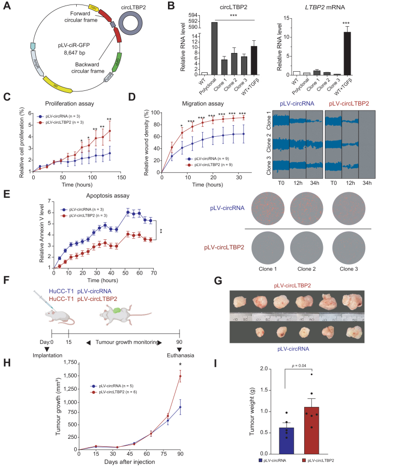
Results: CircLTBP2 (has_circ_0032603) was identified as a novel TGFβ-induced circRNA in several cholangiocarcinoma cell lines. CircLTBP2 promotes tumour cell proliferation, migration, and resistance to gemcitabine-induced apoptosis in vitro and tumour growth in vivo. Mechanistically, circLTBP2 acts as a competitive RNA regulating notably the activity of the tumour suppressor microRNA miR-338-3p, leading to the overexpression of its pro-metastatic targets. The restoration of miR-338-3p levels in iCCA cells reversed the pro-tumourigenic effects driven by circLTBP2, including the resistance to gemcitabine-induced apoptosis. In addition, circLTBP2 expression predicted a reduced survival, as detected in not only tumour tissues but also serum extracellular vesicles isolated from patients with iCCA.

Conclusions: CircLTBP2 is a novel effector of the pro-tumourigenic arm of TGFβ and a clinically relevant biomarker easily detected from liquid biopsies in iCCA.

Impact and implications: Intrahepatic cholangiocarcinoma (iCCA) is an aggressive cancer with limited therapeutic options. Opening the field to new concepts is urgently needed to improve the survival of patients. Here, we evaluated the role and the clinical relevance of circular RNA. We report that TGFβ-induced circLTBP2 contributes to CCA carcinogenesis and may constitute a clinically relevant prognostic biomarker detected in liquid biopsies.

Keywords: Biomarker; Cholangiocarcinoma; Circular RNA; Extracellular vesicles; Transforming growth factor beta.


Conflict of interest statement

JE received honoraria from MSD, Roche, AstraZeneca, and BMS. The authors have no other relevant affiliations or financial involvement with any organisation or entity with a financial interest in or financial conflict with the subject matter or materials discussed in the manuscript. This includes employment, consultancies, honoraria, stock ownership, or options, expert testimony, grants or patents received or pending, or royalties. Please refer to the accompanying ICMJE disclosure forms for further details.

Figures

None
Graphical abstract
Fig. 1
Fig. 1
CircLTBP2, a TGFβ-induced circRNA, predicts a poor prognosis in patients with iCCA. (A) Western blot analysis of p-SMAD2 and SMAD2 expression in HuCC-T1 iCCA cell line in response to TGFβ (1 ng/ml TGFβ, 16 h). (B) Expression of three well-known TGFβ target genes in HuCC-T1: SERPINE1, SMAD7, and BIRC3. Cells were treated with TGFβ (1 ng/ml) alone or in combination with LY2157299 (galunisertib) (10 μM) for 16 h. (C) Hierarchical clustering and volcano plot of circRNA isoforms differentially expressed by TGFβ (1 ng/ml, 16 h) in HuCC-T1. A specific signature of 119 circRNAs, upregulated (UP, n = 85) or downregulated (DN, n = 34) by TGFβ was identified. Horizontal dashed line on the volcano plot: p <0.001. Vertical dashed lines: FC >2. (D) Gene ontology analysis based on circRNA microarray signature showed that dysregulated genes upon TGFβ treatment are involved in the regulation of cell migration and response to apoptosis. Values of p were calculated using the permutation test. (E) Five TGFβ-induced circRNAs randomly selected and their relative expression, as determined by microarray and q-RT-PCR. (F) CircLTBP2 (has_circ_0032603) relative expression in 20 freshly frozen iCCA tumours from a French national cohort (separated in two groups, namely, circLTBP2low and circLTBP2high, according to their circLTBP2 median expression). (G) Kaplan-Meier plots and log-rank statistics analysis revealed a significant decreased OS and relapse-free survival for patients with iCCA with a high expression of circLTBP2. (H) CircLTBP2 relative expression in HCC and CCA tumours unravelled circLTBP2 specificity for CCA. (I) Kaplan-Meier plots and log-rank statistics analysis endorsed the association of circLTBP2 expression with OS in the independent cohort of CCA, whereas no correlation was found in HCC. Statistical analyses for (A–C) were performed using a Mann-Whitney U test. Data are presented as mean ± SD. ∗p <0.05, ∗∗p <0.01, ∗∗∗p<0.001, n ≥3 technical and biological replicates. CCA, cholangiocarcinoma; circRNA, circular RNA; FC, fold change; FDR, false discovery rate; HCC, hepatocellular carcinoma; iCCA, intrahepatic CCA; LTBP2, latent TGFβ-binding protein 2; OS, overall survival; q-RT-PCR, quantitative reverse-transcription PCR; TGFβ, transforming growth factor β.
Fig. 2
Fig. 2
Overexpression of circLTBP2 promotes iCCA cell proliferation, migration, and resistance to gemcitabine-induced apoptosis in vitro and tumour growth in vivo. (A) Schematic representation of circLTBP2 overexpression vector. (B) Relative expression of circLTBP2 and its cognate mRNA in HuCC-T1 clones infected with circLTBP2 overexpression vector. (C) Relative confluency of cultured HuCC-T1 cells was evaluated for 132 h (n = 3 per group). HuCC-T1 cells overexpressing circLTBP2 showed an increased proliferation. (D) Cell migration was evaluated by wound healing assay performed on HuCC-T1 cells overexpressing circLTBP2 compared with the empty vector control group (n = 9 per group). Relative wound density was assessed every 4 h as a percentage of the initial wound surface, represented in blue on the right panel. HuCC-T1 cells were cultured in low FBS concentration medium (1% FBS) and with mitomycin C (10 μg/ml) to inhibit cell proliferation. (E) Resistance of HuCC-T1 cells to gemcitabine-induced apoptosis (10 ng/ml) was evaluated every 4 h using a red fluorescent Annexin V labelling, in both the circLTBP2 overexpressing and control groups. (F) Mouse subcutaneous tumour model was used to evaluate the impact of circLTBP2 overexpression on tumour growth (G, H) and weight (I). Five mice were included in the control group, and 6 mice were included in the circLTBP2 overexpression group. Statistical analyses for (B) and (I) were performed using a Mann-Whitney U test. Statistical analyses for (C–E), and (H) were performed using a two-way ANOVA. ∗p <0.05, ∗∗p <0.01, ∗∗∗p <0.001, n ≥3 technical and biological replicates. CCA, cholangiocarcinoma; circRNA, circular RNA; iCCA, intrahepatic CCA; pLV-circLTBP2, circLTBP2 overexpression vector; pLV-circRNA, empty vector; LTBP2, latent TGFβ-binding protein 2; TGFβ, transforming growth factor β.
Fig 3
Fig 3
CircLTBP2 acts as a sponge for miR-338-3p, a known tumour suppressor miRNA. (A) Schematic representation of the strategy used to select a miRNA showing strong interactions with circLTBP2, based on the data obtained by pulldown assay followed by a miRNA sequencing. (B) Relevance rank of circLTBP2 binding miRNAs based on the predicted number of binding sites (https://starbase.sysu.edu.cn/) and the GSE score of their mRNA targets in the profile of HuCC-T1 cells treated with TGFβ (GSE102109). (C) GSE score of miR-338-3p mRNA targets in HuCC-T1 TGFβ signature (GSE102109). The top 20 enriched genes are listed. (D) RNA IP of AGO2 in HuCC-T1 cells transfected with miR-338-3p mimics and quantification by q-RT-PCR of putative miR-338-3p binding RNA (i.e. circLTBP2). (E) Expression of miR-338-3p targets after TGFβ treatment evaluated by q-RT-PCR. (F) Expression of miR-338-3p targets evaluated by q-RT-PCR in HuCC-T1 cells transfected with miR-338-3p mimics (5 nM). (G) Mir-338-3p relative expression in 20 freshly frozen iCCA tumours from a French national cohort (separated in two groups, namely, miR-338-3plow and miR-338-3phigh, according to their median expression). (H) Kaplan-Meier plots and log-rank statistics analysis of overall and relapse-free survival for patients with iCCA with high vs. low expression of miR-338-3p. Statistical analyses for (D–G) were performed using a Mann-Whitney U test. ∗p <0.05, ∗∗p <0.01, ∗∗∗p <0.001, n ≥3 technical and biological replicates. CCA, cholangiocarcinoma; GSEA, gene set enrichment analysis; iCCA, intrahepatic CCA; IP, immunoprecipitation; LTBP2, latent TGFβ-binding protein 2; miRNA, microRNA; NES, normal enrichment score; q-RT-PCR, quantitative reverse-transcription PCR; TGFβ, transforming growth factor β.
Fig. 4
Fig. 4
The restoration of miR-338-3p levels in iCCA cells reversed the pro-tumourigenic effects driven by circLTBP2. (A) Relative levels of miR-338-3p in HuCC-T1 were assessed by q-RT-PCR in cells overexpressing circLTBP2 vs. cells infected with the empty vector. In addition, miR-338-3p mimics (5 nM) were added with the aim of rescuing the sponging mediated by circLTBP2. (B) Relative confluency of cultured HuCC-T1 cells was evaluated for 96 h in HuCC-T1 cells overexpressing circLTBP2 compared with the empty vector control group (n = 3 per group). In addition, miR-338-3p mimics were added aiming at restoring the phenotype. (C) Cell migration was evaluated by wound healing assay performed on HuCC-T1 cells overexpressing circLTBP2 compared with the empty vector control group (n = 3 per group). In addition, miR-338-3p mimics (5 nM) were added aiming at restoring the phenotype. (D) Resistance of HuCC-T1 cells to gemcitabine-induced apoptosis (10 ng/ml) was evaluated every 4 h using a red fluorescent Annexin V labelling, in both the circLTBP2 overexpressing and control groups. In addition, miR-338-3p mimics were added aiming at restoring the phenotype (5 nM). Statistical analyses for (A) were performed using a Mann-Whitney U test. Statistical analyses for (B–D) were performed using a two-way ANOVA. ∗p <0.05, ∗∗p <0.01, ∗∗∗p <0.001, n ≥3 technical and biological replicates. CCA, cholangiocarcinoma; circRNA, circular RNA; iCCA, intrahepatic CCA; pLV-circLTBP2, circLTBP2 overexpression vector; pLV-circRNA, empty vector; LTBP2, latent TGFβ-binding protein 2; q-RT-PCR, quantitative reverse-transcription PCR; TGFβ, transforming growth factor β.
Fig. 5
Fig. 5
Circulating vesicles containing circLTBP2 in serum are associated with poor OS in patients with iCCA. (A) Size and concentration of EVs collected from HuCC-T1 supernatant by UC were assessed using a tunable resistive sensing method (qNano, Izon Science). EVs with a size ranging from 100 to 200 nm were characterised. (B) Scanning electron microscopy was used to image EVs collected after UC. The morphology and size observed match those of EVs. (C) Immunoblotting of EV marker CD9 in HuCC-T1 cells and EVs isolated from their supernatant. The protein GRP78 was used as a cell specific negative control. (D) Relative levels of circLTBP2 in HuCC-T1 cells and EVs isolated from their supernatant by UC. HuCC-T1 cells were first treated with TGFβ (1 ng/ml, 16 h). (E) Half-life of circLTBP2 in HuCC-T1 cells treated with actinomycin D (2 μg/ml) for 24 h. (F) Quantification by q-RT-PCR of EV-embed circLTBP2 in sera from patients with metastatic advanced iCCA (Rennes cohort, n = 62). Sera from healthy individuals were used as control (n = 12). (G) Positive correlation of circLTBP2 expression in matched tissues and serum samples (patients for which both samples are available, n = 17). (H) EV-embed circLTBP2 relative expression in sera from patients with iCCA whose survival data were available (separated in two groups, namely, circLTBP2low and circLTBP2high, according to their circLTBP2 median expression). (I) Kaplan-Meier plots and log-rank statistics analysis revealed a significant decreased OS for patients with iCCA with a high expression of vesicles-embed circLTBP2. Levels of circLTBP2 were not associated with the RFS. Statistical analyses for (A) were performed using a Mann-Whitney U test. Statistical analyses for (B–D) were performed using a two-way ANOVA. ∗p <0.05, ∗∗p <0.01, ∗∗∗p <0.001, n ≥3 technical and biological replicates. CCA, cholangiocarcinoma; EV, extracellular vesicle; iCCA, intrahepatic CCA; LTBP2, latent TGFβ-binding protein 2; OS, overall survival; pLV-circLTBP2, circLTBP2 overexpression vector; pLV-circRNA, empty vector; q-RT-PCR, quantitative reverse-transcription PCR; RFS, relapse-free survival; TGFβ, transforming growth factor β; UC, ultracentrifugation.

References

    1. Banales J.M., Marin J.J.G., Lamarca A., Rodrigues P.M., Khan S.A., Roberts L.R., et al. Cholangiocarcinoma 2020: the next horizon in mechanisms and management. Nat Rev Gastroenterol Hepatol. 2020;17:557–588. - PMC - PubMed
    1. Valle J.W., Kelley R.K., Nervi B., Oh D.Y., Zhu A.X. Biliary tract cancer. Lancet. 2021;397:428–444. - PubMed
    1. Izquierdo-Sanchez L., Lamarca A., La Casta A., Buettner S., Utpatel K., Klüumpen H.J., et al. Cholangiocarcinoma landscape in Europe: diagnostic, prognostic and therapeutic insights from the ENSCCA Registry. J Hepatol. 2022;76:1109–1121. - PubMed
    1. Macias R.I.R., Cardinale V., Kendall T.J., Avila M.A., Guido M., Coulouarn C., et al. Clinical relevance of biomarkers in cholangiocarcinoma: critical revision and future directions. Gut. 2022;71:1669–1683. - PubMed
    1. Rizvi S., Khan S.A., Hallemeier C.L., Kelley R.K., Gores G.J. Cholangiocarcinoma – evolving concepts and therapeutic strategies. Nat Rev Clin Oncol. 2018;15:95–111. - PMC - PubMed