. 2023 Nov 9;5(1):vdad149.
doi: 10.1093/noajnl/vdad149. eCollection 2023 Jan-Dec.

Longitudinal characteristics of T2-FLAIR mismatch in IDH-mutant astrocytomas: Relation to grade, histopathology, and overall survival in the GLASS-NL cohort

Affiliations

Longitudinal characteristics of T2-FLAIR mismatch in IDH-mutant astrocytomas: Relation to grade, histopathology, and overall survival in the GLASS-NL cohort

Karin A van Garderen et al. Neurooncol Adv. .

Abstract

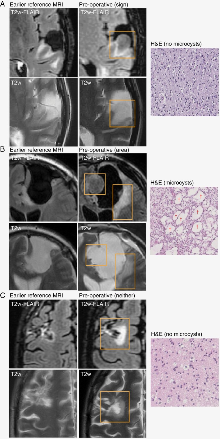
Background: The T2-FLAIR mismatch sign is defined by signal loss of the T2-weighted hyperintense area with Fluid-Attenuated Inversion Recovery (FLAIR) on magnetic resonance imaging, causing a hypointense region on FLAIR. It is a highly specific diagnostic marker for IDH-mutant astrocytoma and is postulated to be caused by intercellular microcystic change in the tumor tissue. However, not all IDH-mutant astrocytomas show this mismatch sign and some show the phenomenon in only part of the lesion. The aim of the study is to determine whether the T2-FLAIR mismatch phenomenon has any prognostic value beyond initial noninvasive molecular diagnosis.

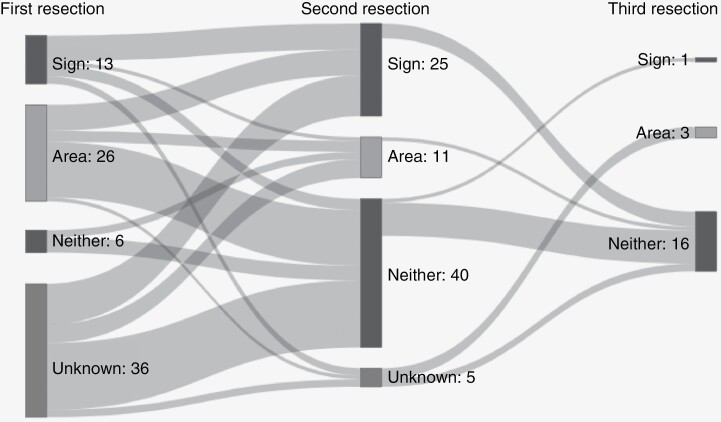
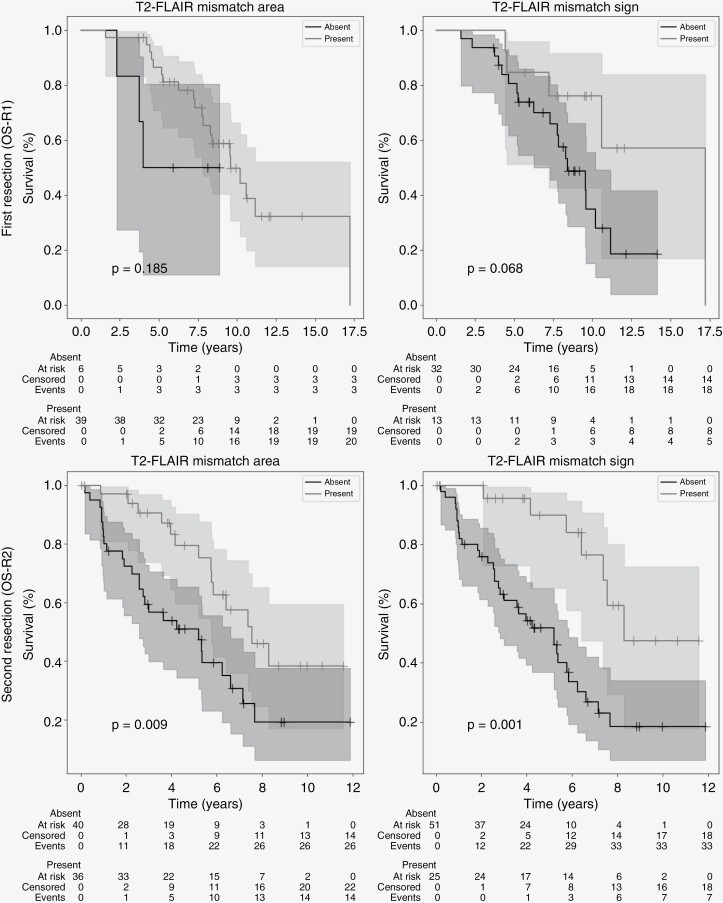
Methods: Patients initially diagnosed with histologically lower-grade (2 or 3) IDH-mutant astrocytoma and with at least 2 surgical resections were included in the GLASS-NL cohort. T2-FLAIR mismatch was determined, and the growth pattern of the recurrent tumor immediately before the second resection was annotated as invasive or expansive. The relation between the T2-FLAIR mismatch sign and tumor grade, microcystic change, overall survival (OS), and other clinical parameters was investigated both at first and second resection.

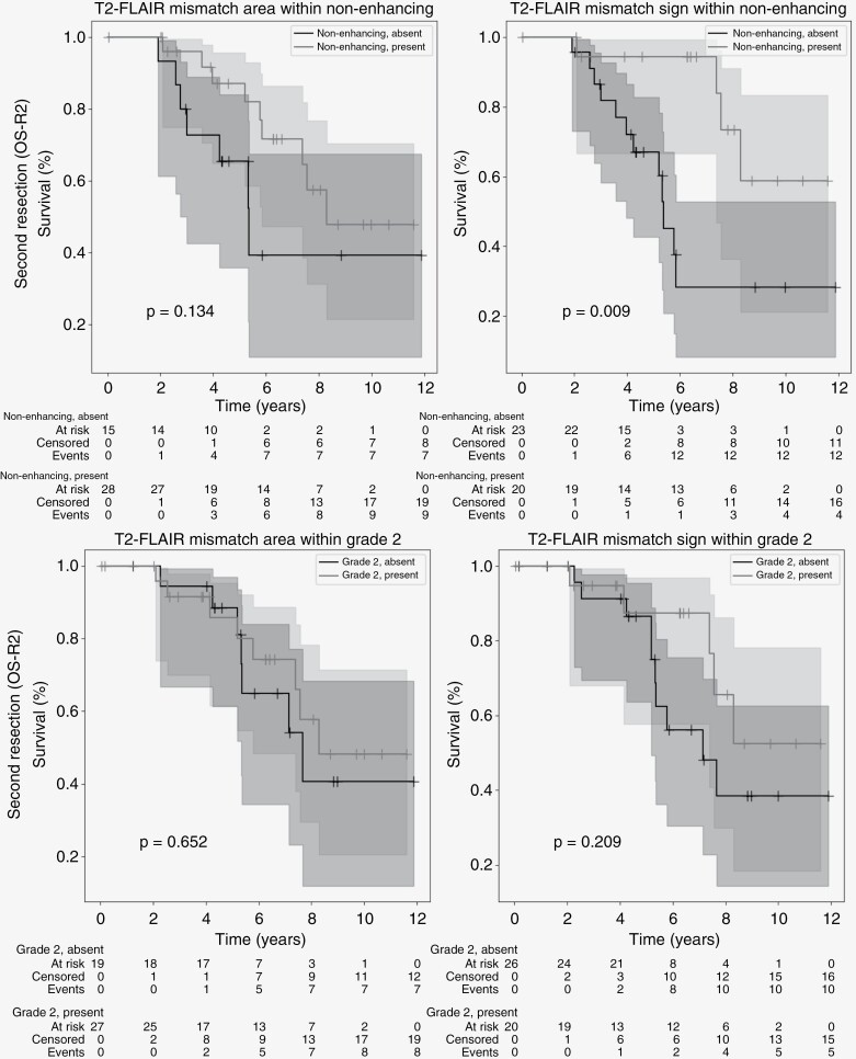
Results: The T2-FLAIR mismatch sign was significantly related to Grade 2 (80% vs 51%), longer post-resection median OS (8.3 vs 5.2 years), expansive growth, and lower age at second resection. At first resection, no relation was found between the mismatch sign and OS. Microcystic change was associated with areas of T2-FLAIR mismatch.

Conclusions: T2-FLAIR mismatch in IDH-mutant astrocytomas is correlated with microcystic change in the tumor tissue, favorable prognosis, and Grade 2 tumors at the time of second resection.

Keywords: astrocytoma; glioma; magnetic resonance imaging.


Conflict of interest statement

M.S. reports receiving speaker fees (paid to the institution) from GE Health care and AuntMinnie.com, and consulting fees (paid to the institution) from Bracco SpA. The other authors declare no conflicts of interest.

Figures

Figure 1.
Figure 1.
Examples of FLAIR and T2w MRI and H&E slides for 3 cases. The last preoperative MRI is shown with an earlier reference MRI used to assess the growth pattern. Recurrent lesions are outlined with a rectangle. Examples of microcysts are indicated by a “†” symbol. (A) Recurrent tumor with mismatch sign; preoperative MRI shows pre-existing treatment effect, but recurrent growth is mismatched with hyperintense rim; growth pattern is mostly expansive; H&E does not show microcysts. (B) Recurrent tumor that is partly mismatched, so this case shows a T2-FLAIR mismatch area but no mismatch sign; growth pattern is mixed; H&E shows large microcysts. Note that the source of the histopathology (mismatch area or not) is unknown. (C) Recurrent tumor without T2-FLAIR mismatch; growth pattern is mostly invasive; H&E shows no microcysts.
Figure 2.
Figure 2.
Sankey diagram of T2-FLAIR mismatch sign and area over repeated resections (left: first, middle: second, and right: third). Area indicates that there was an area of T2-FLAIR mismatch, but the lesion did not meet the criteria for the mismatch sign. Resections of the same patients are connected by gray bands.
Figure 3.
Figure 3.
Kaplan–Meier curves for OS-R2 of mismatch area (left) and mismatch sign (right) at first (top) and second (bottom) resection. Starting date for the analysis is the date of first and second resection, respectively, and T2-FLAIR mismatch area/sign was annotated on the last available MRI before resection. Censored patients indicated by a “+” at date of last follow-up. Shaded areas indicate 95% confidence intervals.
Figure 4.
Figure 4.
Kaplan–Meier curves of mismatch area (left) and sign (right) combined with other indicators of good prognosis at second resection. Top: Nonenhancing lesions. Bottom: WHO-2021 Grade 2. Starting date of the analysis is date of second resection. T2-FLAIR mismatch area/sign and enhancement were annotated on the last available MRI before resection. Censored patients indicated by a “+” at date of last follow-up. Shaded areas indicate 95% confidence intervals.

References

    1. Aldape K, Amin SB, Ashley DM, et al. . Glioma through the looking GLASS: molecular evolution of diffuse gliomas and the Glioma Longitudinal Analysis Consortium. Neuro-Oncol. 2018;20(7):873–884. - PMC - PubMed
    1. Patel SH, Poisson LM, Brat DJ, et al. . T2–FLAIR mismatch, an imaging biomarker for IDH and 1p/19q status in lower-grade gliomas: a TCGA/TCIA project. Clin Cancer Res. 2017;23(20):6078–6085. - PubMed
    1. Corell A, Ferreyra Vega S, Hoefling N, et al. . The clinical significance of the T2-FLAIR mismatch sign in grade II and III gliomas: a population-based study. BMC Cancer. 2020;20(1):1–10. - PMC - PubMed
    1. Broen MPG, Smits M, Wijnenga MMJ, et al. . The T2-FLAIR mismatch sign as an imaging marker for non-enhancing IDH-mutant, 1p/19q-intact lower-grade glioma: a validation study. Neuro Oncol. 2018;20(10):1393–1399. - PMC - PubMed
    1. Deguchi S, Oishi T, Mitsuya K, et al. . Clinicopathological analysis of T2-FLAIR mismatch sign in lower-grade gliomas. Sci Rep. 2020;10(1):111–116. - PMC - PubMed

LinkOut - more resources