

Diffusion in the corpus callosum predicts persistence of clinical symptoms after mild traumatic brain injury, a multi-scanner study
- PMID: 38025312
- PMCID: PMC10654678
- DOI: 10.3389/fnimg.2023.1153115
Diffusion in the corpus callosum predicts persistence of clinical symptoms after mild traumatic brain injury, a multi-scanner study
Abstract
Background: Mild traumatic brain injuries (mTBIs) comprise 80% of all TBI, but conventional MRI techniques are often insensitive to the subtle changes and injuries produced in a concussion. Diffusion tensor imaging (DTI) is one of the most sensitive MRI techniques for mTBI studies with outcome and symptom associations described. The corpus callosum (CC) is one of the most studied fiber tracts in TBI and mTBI, but the comprehensive post-mTBI symptom relationship has not fully been explored.
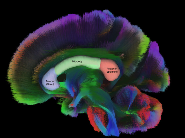
Methods: This is a retrospective observational study of how quantitative DTI data of the CC and its sub-regions may relate to clinical presentation of symptoms and timing of resolution of symptoms in patients diagnosed with uncomplicated mTBI. DTI and clinical data were obtained retrospectively from 446 (mean age 42 years, range 13-82) civilian patients. From patient medical charts, presentation of the following common post-concussive symptoms was noted: headache, balance issues, cognitive deficits, fatigue, anxiety, depression, and emotional lability. Also recorded was the time between injury and a visit to the physician when improvement or resolution of a particular symptom was reported. FA values from the total CC and 3 subregions of the CC (genu or anterior, mid body, and splenium or posterior) were obtained from hand tracing on the Olea Sphere v3.0 SP12 free-standing workstation. DTI data was obtained from 8 different 3T MRI scanners and harmonized via ComBat harmonization. The statistical models used to explore the association between regional Fractional Anisotropy (FA) values and symptom presentation and time to symptom resolution were logistic regression and interval-censored semi-parametric Cox proportional hazard models, respectively. Subgroups related to age and timing of first scan were also analyzed.
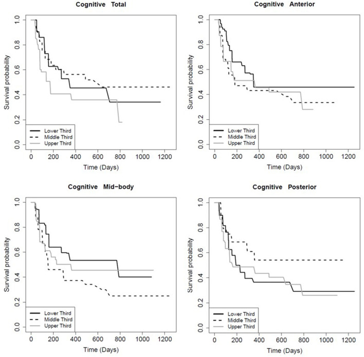
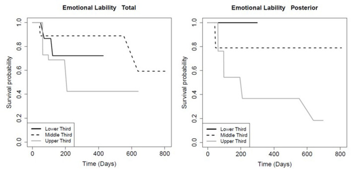
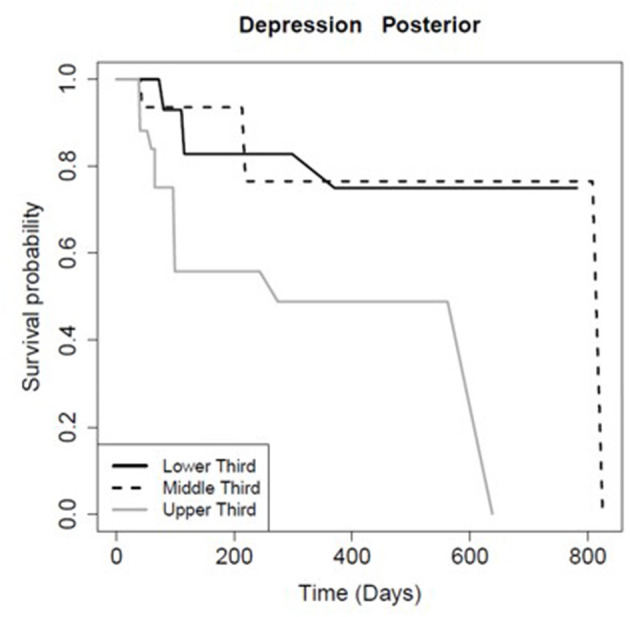
Results: Patients with the highest FA in the total CC (p = 0.01), anterior CC (p < 0.01), and mid-body CC (p = 0.03), but not the posterior CC (p = 0.91) recovered faster from post-concussive cognitive deficits. Patients with the highest FA in the posterior CC recovered faster from depression (p = 0.04) and emotional lability (p = 0.01). There was no evidence that FA in the CC or any of its sub-regions was associated with symptom presentation or with time to resolution of headache, balance issues, fatigue, or anxiety. Patients with mTBI under 40 had higher FA in the CC and the anterior and mid-body subregions (but not the posterior subregion: p = 1.00) compared to patients 40 or over (p ≤ 0.01). There was no evidence for differences in symptom presentation based on loss of consciousness (LOC) or sex (p ≥ 0.18).
Conclusion: This study suggests that FA of the CC has diagnostic and prognostic value for clinical assessment of mTBI in a large diverse civilian population, particularly in patients with cognitive symptoms.
Keywords: clinical outcomes; concussion; diffusion tensor imaging (DTI); interval-censored; post-concussion syndrome (PCS).
Copyright © 2023 Asturias, Knoblauch, Rodriguez, Vanier, Le Tohic, Barrett, Eisenberg, Gibbert, Zimmerman, Parikh, Nguyen, Azad, Germin, Fazzini and Snyder.
Conflict of interest statement
SA and TS were employed by company HCA Healthcare. AA, TK, AR, CV, and TS were employed by company Imgen Research Group. LG was employed by company Clinical Neurology Specialists. TS was employed by company SimonMed Imaging. The remaining authors declare that the research was conducted in the absence of any commercial or financial relationships that could be construed as a potential conflict of interest.
Figures
References
-
- American Psychiatric Association (2013). Diagnostic and Statistical Manual of Mental Disorders. Washington DC: American Psychiatric Association.
LinkOut - more resources
Full Text Sources