. 2023 Nov 14:11:e16236.
doi: 10.7717/peerj.16236. eCollection 2023.

Osteology of the axial skeleton of Aucasaurus garridoi: phylogenetic and paleobiological inferences

Affiliations

Osteology of the axial skeleton of Aucasaurus garridoi: phylogenetic and paleobiological inferences

Mattia Antonio Baiano et al. PeerJ. .

Abstract

Aucasaurus garridoi is an abelisaurid theropod from the Anacleto Formation (lower Campanian, Upper Cretaceous) of Patagonia, Argentina. The holotype of Aucasaurus garridoi includes cranial material, axial elements, and almost complete fore- and hind limbs. Here we present a detailed description of the axial skeleton of this taxon, along with some paleobiological and phylogenetic inferences. The presacral elements are somewhat fragmentary, although these show features shared with other abelisaurids. The caudal series, to date the most complete among brachyrostran abelisaurids, shows several autapomorphic features including the presence of pneumatic recesses on the dorsal surface of the anterior caudal neural arches, a tubercle lateral to the prezygapophysis of mid caudal vertebrae, a marked protuberance on the lateral rim of the transverse process of the caudal vertebrae, and the presence of a small ligamentous scar near the anterior edge of the dorsal surface in the anteriormost caudal transverse process. The detailed study of the axial skeleton of Aucasaurus garridoi has also allowed us to identify characters that could be useful for future studies attempting to resolve the internal phylogenetic relationships of Abelisauridae. Computed tomography scans of some caudal vertebrae show pneumatic traits in neural arches and centra, and thus the first reported case for an abelisaurid taxon. Moreover, some osteological correlates of soft tissues present in Aucasaurus and other abelisaurids, especially derived brachyrostrans, underscore a previously proposed increase in axial rigidity within Abelisauridae.

Keywords: Abelisauridae; Anacleto formation; Brachyrostra; Late Cretaceous; Patagonia; Phylogeny; Pneumaticity; Theropoda.


Conflict of interest statement

Luis Chiappe is an Academic Editor for PeerJ.

Figures

Figure 1
Figure 1. Axial skeleton of Aucasaurus garridoi.
Lateral right view of the axial elements of the holotype MCF-PVPH-236. Scale bar: 1 m. Silhouette modified from Scott Hartman (https://www.skeletaldrawing.com).
Figure 2
Figure 2. Atlas of Aucasaurus garridoi MCF-PVPH-236.
In anterior (A), posterior (B), right lateral (C), ventral (D), and dorsal (E) views. amp, anteromedial process; ic, intercentrum; nrp, neurapophysis; od, odontoid; vp, ventral process. Scale bar: 5 cm.
Figure 3
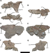
Figure 3. Cervical vertebra fragments of Aucasaurus garridoi MCF-PVPH-236.
In lateral (A, G and E), ventral (B), dorsal (C), and medial (D and F) views. ape, anterior process of epipophysis; eprl, epipophyseal prezygapophyseal lamina; podl, postzygodiapophyseal lamina; poz, postzygapophysis; ppe, posterior process of epipophysis; prz, prezygapophysis; sprl, spinoprezigapophyseal lamina; tp, transverse process. Scale bar: 5 cm.
Figure 4
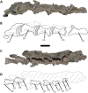
Figure 4. (A–B) Photographs and line drawings of the anterior dorsal vertebrae of Aucasaurus garridoi MCF-PVPH-236.
In lateral (A) view. 2dns, second dorsal neural spine; 7dns, seventh dorsal neural spine; acpl, anterior centroparapophyseal lamina; D2–D6, second to seventh dorsal vertebrae; iap, interspinous accessory process; ilp, interspinous ligament process; pl, pleurocoel; pp, parapophysis; prz, prezygapophysis; tp, transverse process. Scale bar: 5 cm.
Figure 5
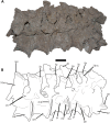
Figure 5. Photographs and line drawings of the anterior dorsal vertebrae of Aucasaurus garridoi MCF-PVPH-236.
In ventral (A and B), and dorsal (C and D) views. Abbreviations: D2–D7, second to seventh dorsal vertebrae; iap, interspinous accessory process; ilp, interspinous ligament process. Scale bar: 5 cm.
Figure 6
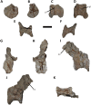
Figure 6. Posterior dorsal vertebrae of Aucasaurus garridoi MCF-PVPH-236.
In anterior (A and G), posterior (B and H), lateral (C, D, I and J), dorsal (E), and ventral (F and K) views. ns, neural spine; pl, pleurocoel. Scale bar: 5 cm.
Figure 7
Figure 7. Distal fragments of dorsal neural spines of Aucasaurus garridoi MCF-PVPH-236.
In dorsal (A–C), and left lateral (D–F) views. iap, interspinous accessory process; ilp, interspinous ligament process. Scale bar: 5 cm.
Figure 8
Figure 8. Sacrum of Aucasaurus garridoi MCF-PVPH-236.
In lateral (A and B), ventral (C), posterior (D), and dorsal (E and F) views. Colored dashed lines marking the anterior and posterior rims of the third to fifth transverse processes. 1sc–6sc, first to sixth sacral centra; 4sr, fourth sacral rib; 1stp–5stp, first to fifth sacral transverse processes; IL, ilion; ns, neural spine. Scale bar: 10 cm.
Figure 9
Figure 9. First caudal vertebra of Aucasaurus garridoi MCF-PVPH-236.
In anterior (A), lateral (B and E), dorsal (C), posterior (D), and ventral (F) views. acdl, anterior centrodiapophyseal lamina; apltp, anterior process of lateral transverse process; cdl, centrodiapophyseal lamina; dr, dorsal roughness; ha, hypantrum; hy, hyposphene; iap, interspinous accessory process; ldvc, lateral depression of vertebral centrum; lrcdl, lateral ridge of centrodiapophyseal lamina; nc, neural canal; ns, neural spine; pcdl, posterior centrodiapophyseal lamina; pf, pneumatic foramen; poz, postzygapophysis; prz, prezygapophysis; spof, spinopostzigapophyseal fossa; spol, spinopostzigapophyseal lamina; sprf, spinoprezigapophyseal fossa; tp, transverse process; vlrtp, ventrolateral ridge of the transverse process. Scale bar: 10 cm.
Figure 10
Figure 10. Second caudal vertebra of Aucasaurus garridoi MCF-PVPH-236.
In anterior (A), lateral (B and E), dorsal (C), posterior (D), and ventral (F) views. acdl, anterior centrodiapophyseal lamina; apltp, anterior process of lateral transverse process; cdl, centrodiapophyseal lamina; ha, hypantrum; haaf, haemal arch articular facet; hy, hyposphene; ldvc, lateral depression of vertebral centrum; lrcdl, lateral ridge of centrodiapophyseal lamina; nc, neural canal; ns, neural spine; pcdl, posterior centrodiapophyseal lamina; pf, pneumatic foramen; poz, postzygapophysis; ppltp, posterior process of lateral transverse process; prz, prezygapophysis; spof, spinopostzigapophyseal fossa; spol, spinopostzigapophyseal lamina; sprf, spinoprezigapophyseal fossa; tp, transverse process; vg, ventral groove; vlrtp, ventrolateral ridge of the transverse process. Scale bar: 10 cm.
Figure 11
Figure 11. Third caudal vertebra of Aucasaurus garridoi MCF-PVPH-236.
In anterior (A), lateral (B and E), dorsal (C), posterior (D), and ventral (F) views. acdl, anterior centrodiapophyseal lamina; apltp, anterior process of lateral transverse process; cdf, centrodiapophyseal fossa; cdl, centrodiapophyseal lamina; dr, dorsal roughness; ha, hypantrum; haaf, haemal arch articular facet; hy, hyposphene; ldvc, lateral depression of vertebral centrum; lrcdl, lateral ridge of centrodiapophyseal lamina; lrtp, lateral rugosity of transverse process; nc, neural canal; ns, neural spine; pcdl, posterior centrodiapophyseal lamina; pocdf, postzygapophyseal centrodiapophyseal fossa; poz, postzygapophysis; prcdf, prezygapophyseal centrodiapophyseal fossa; prz, prezygapophysis; spof, spinopostzigapophyseal fossa; spol, spinopostzigapophyseal lamina; sprf, spinoprezigapophyseal fossa; vg, ventral groove; vlrtp, ventrolateral ridge of the transverse process. Scale bar: 10 cm.
Figure 12
Figure 12. Fourth caudal vertebra of Aucasaurus garridoi MCF-PVPH-236.
In anterior (A), lateral (B and E), dorsal (C), posterior (D), and ventral (F) views. acdl, anterior centrodiapophyseal lamina; apltp, anterior process of lateral transverse process; cdf, centrodiapophyseal fossa; cdl, centrodiapophyseal lamina; dr, dorsal roughness; haaf, haemal arch articular facet; hy, hyposphene; ldvc, lateral depression of vertebral centrum; lrcdl, lateral ridge of centrodiapophyseal lamina; lrtp, lateral rugosity of transverse process; nc, neural canal; ns, neural spine; pcdl, posterior centrodiapophyseal lamina; pocdf, postzygapophyseal centrodiapophyseal fossa; poz, postzygapophysis; prcdf, prezygapophyseal centrodiapophyseal fossa; prz, prezygapophysis; spof, spinopostzigapophyseal fossa; spol, spinopostzigapophyseal lamina; sprf, spinoprezigapophyseal fossa; vg, ventral groove. Scale bar: 10 cm.
Figure 13
Figure 13. Fifth and sixth caudal vertebrae of Aucasaurus garridoi MCF-PVPH-236.
In anterior (A), lateral (B and E), dorsal (C), posterior (D), and ventral (F) views. 5 cv, fifth caudal vertebra; 6 cv, sixth caudal vertebra; apltp, anterior process of lateral transverse process; cdl, centrodiapophyseal lamina; dr, dorsal roughness; ha, hypantrum; har, haemal arch; haaf, haemal arch articular facet; hy, hyposphene; iap; interspinous accessory process; lrcdl, lateral ridge of centrodiapophyseal lamina; lrtp, lateral rugosity of transverse process; nc, neural canal; ns, neural spine; pf, pneumatic foramen; poz, postzygapophysis; prcdf, prezygapophyseal centrodiapophyseal fossa; prz, prezygapophysis; spof, spinopostzigapophyseal fossa; spol, spinopostzigapophyseal lamina; sprf, spinoprezigapophyseal fossa; tp, transverse process; vg, ventral groove; vlrtp, ventrolateral ridge of the transverse process. Scale bar: 10 cm.
Figure 14
Figure 14. Seventh caudal vertebra of Aucasaurus garridoi MCF-PVPH-236.
In anterior (A), lateral (B and E), dorsal (C), posterior (D), and ventral (F) views. acdl, anterior centrodiapophyseal lamina; apltp, anterior process of lateral transverse process; cdf, centrodiapophyseal fossa; cdl, centrodiapophyseal lamina; dr, dorsal roughness; ha, hypantrum; haaf, haemal arch articular facet; hy, hyposphene; iap; interspinous accessory process; lrcdl, lateral ridge of centrodiapophyseal lamina; lrtp, lateral rugosity of transverse process; nc, neural canal; ns, neural spine; pcdl, posterior centrodiapophyseal lamina; pf, pneumatic foramen; poz, postzygapophysis; prcdf, prezygapophyseal centrodiapophyseal fossa; prz, prezygapophysis; spof, spinopostzigapophyseal fossa; sprf, spinoprezigapophyseal fossa; sprl, spinoprezigapophyseal lamina; vg, ventral groove. Scale bar: 10 cm.
Figure 15
Figure 15. Eighth caudal vertebra of Aucasaurus garridoi MCF-PVPH-236.
In anterior (A), lateral (B and E), dorsal (C), posterior (D), and ventral (F) views. apbns, anterior process of basal neural spine; apltp, anterior process of lateral transverse process; cdl, centrodiapophyseal lamina; dr, dorsal roughness; haaf, haemal arch articular facet; hy, hyposphene; lrcdl, lateral ridge of centrodiapophyseal lamina; lrtp, lateral rugosity of transverse process; nc, neural canal; ns, neural spine; pocdf, postzygapophyseal centrodiapophyseal fossa; poz, postzygapophysis; prcdf, prezygapophyseal centrodiapophyseal fossa; prz, prezygapophysis; spof, spinopostzigapophyseal fossa; spol, spinopostzigapophyseal lamina; sprf, spinoprezigapophyseal fossa; sprl, spinoprezigapophyseal lamina; vg, ventral groove. Scale bar: 10 cm.
Figure 16
Figure 16. Ninth caudal vertebra of Aucasaurus garridoi MCF-PVPH-236.
In anterior (A), lateral (B and E), dorsal (C), posterior (D), and ventral (F) views. apltp, anterior process of lateral transverse process; cdl, centrodiapophyseal lamina; haaf, haemal arch articular facet; hy, hyposphene; lrtp, lateral rugosity of transverse process; nc, neural canal; ns, neural spine; pf, pneumatic foramen; pocdf, postzygapophyseal centrodiapophyseal fossa; poz, postzygapophysis; prcdf, prezygapophyseal centrodiapophyseal fossa; prz, prezygapophysis; spof, spinopostzigapophyseal fossa; spol, spinopostzigapophyseal lamina; sprf, spinoprezigapophyseal fossa; sprl, spinoprezigapophyseal lamina; vg, ventral groove. Scale bar: 10 cm.
Figure 17
Figure 17. Tenth caudal vertebra of Aucasaurus garridoi MCF-PVPH-236.
In anterior (A), lateral (B and E), dorsal (C), posterior (D), and ventral (F) views. apltp, anterior process of lateral transverse process; cdl, centrodiapophyseal lamina; dr, dorsal roughness; haaf, haemal arch articular facet; lrcdl, lateral ridge of centrodiapophyseal lamina; lrtp, lateral rugosity of transverse process; nc, neural canal; ns, neural spine; pf, pneumatic foramen; prcdf, prezygapophyseal centrodiapophyseal fossa; prz, prezygapophysis; sprf, spinoprezigapophyseal fossa. Scale bar: 10 cm.
Figure 18
Figure 18. Eleventh caudal vertebra of Aucasaurus garridoi MCF-PVPH-236.
In anterior (A), lateral (B and E), dorsal (C), posterior (D), and ventral (F) views. apltp, anterior process of lateral transverse process; dr, dorsal roughness; haaf, haemal arch articular facet; lrtp, lateral rugosity of transverse process; nc, neural canal; ns, neural spine; pf, pneumatic foramen; poz, postzygapophysis; prz, prezygapophysis; vg, ventral groove. Scale bar: 10 cm.
Figure 19
Figure 19. Twelfth and thirteenth caudal vertebrae of Aucasaurus garridoi MCF-PVPH-236.
In anterior (A), lateral (B and E), dorsal (C), posterior (D), and ventral (F) views. 12 cv, twelfth posterior vertebra; 13 cv, thirteenth posterior vertebra; apltp, anterior process of lateral transverse process; dr, dorsal roughness; haaf, haemal arch articular facet; ltprz, lateral tubercle of prezygapophysis; lrtp, lateral rugosity of transverse process; nc, neural canal; ns, neural spine; poz, postzygapophysis; prz, prezygapophysis; vg, ventral groove. Scale bar: 10 cm.
Figure 20
Figure 20. Caudal neural spines of Aucasaurus garridoi MCF-PVPH-236.
In lateral (A and B) and dorsal (C and D) views. iap, interspinous accessory process. Scale bar: 5 cm.
Figure 21
Figure 21. Caudal transverse processes of Aucasaurus garridoi MCF-PVPH-236.
In dorsal (A and B) and ventral (C and D) views. Abbreviations: apltp, anterior process of lateral transverse process; cdl, centrodiapophyseal lamina; dr, dorsal roughness; lrcdl, lateral ridge of centrodiapophyseal lamina; lrtp, lateral rugosity of transverse process. Scale bar: 5 cm.
Figure 22
Figure 22. Proximal fragments of two cervical ribs of Aucasaurus garridoi MCF-PVPH-23.
In lateral (A and B) views. alp, anterolateral process; cap, capitulum; dlp, dorsolateral process; tub, tuberculum. Scale bar: 5 cm.
Figure 23
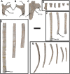
Figure 23. Fragments of dorsal ribs of Aucasaurus garridoi MCF-PVPH-236.
In lateral (A and C–G) and medial (B) views. cap, capitulum; ctw, capitotuberculum web; der, distal expansion of rib; drcap, distal ridge of capitulum; ir, intercostal ridge; tub, tuberculum. Scale bar: 5 cm.
Figure 24
Figure 24. Gastralia of Aucasaurus garridoi MCF-PVPH-236.
In ventral (A, C, and D) and dorsal (B) views. dpg, distal process of gastralia; g, groove; mfg, medial fusion of gastralia; mpg, medial process of the gastralium. Scale bar: 5 cm.
Figure 25
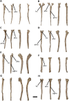
Figure 25. First to eighth haemal arches of Aucasaurus garridoi MCF-PVPH-236.
In anterior, posterior, and lateral (A–H; from left to right) views. afcc, articular facet for the caudal centrum; arha, anterior ridge of haemal arch; hc, haemal canal; pgha, posterior groove of the haemal arch; prha, posterior ridge of the haemal arch. Scale bar: 5 cm.
Figure 26
Figure 26. Ninth to thirteenth haemal arches of Aucasaurus garridoi MCF-PVPH-236.
In anterior, posterior, and lateral (A–E; from left to right) views. hc, haemal canal; pgha, posterior groove of the haemal arc. Scale bar: 5 cm.
Figure 27
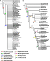
Figure 27. Phylogenetic relationships of Aucasaurus garridoi MCF-PVPH-236.
The results show a quite unresolved strisct consensus (A), and a more resolved topology of the reduced consensus. (B) Colored dots were used for node-based taxa, colored arrows for stem-based taxa.
Figure 28
Figure 28. Photographs of autapomorphies of Aucasaurus garridoi.
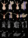
Dorsal view of the odontoids of Aucasaurus (A), Carnotaurus (B), Thanos (C), Majungasaurus (modified from O’Connor, 2007) (E), and Masiakasaurus (modified from Carrano, Loewen & Sertich, 2011) (D). Image not to scale.
Figure 29
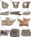
Figure 29. Photographs of autapomorphies of Aucasaurus garridoi.
Outline (in red dashed line) of the anterior articular surface of the atlas of Aucasaurus (A), Viavenator (B), and Carnotaurus (C). Interspinous accessory processes on the dorsal (D), sacral (E), and caudal (F) neural spines of Aucasaurus. Lateral tubercle of prezygapophysis in the middle caudal vertebrae of Aucasaurus (G), and ventral bulge on prezygapophysis of the posterior caudal vertebrae of Alioramus (modified from Brusatte, Carr & Norell, 2012) (H). Foramina on the dorsal surface of the caudal neural arch in Meraxes (I), whereas Aucasaurus holds pneumatic foramina on the dorsal surface of the neural arches (framed by blue dashed lines) of the ninth (J) and eleventh (K) caudal vertebrae. Image not to scale.
Figure 30
Figure 30. Photographs of autapomorphies of Aucasaurus garridoi.
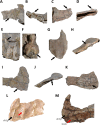
Lateral rugosity and tubercle of the transverse processes of the fourth (A), ninth (B), and eleventh (C) caudal vertebrae of Aucasaurus in dorsal (upper) and lateral (lower) views. Anterodorsal scar (black arrows) of the transverse processes of the first (D), and second (E) caudal vertebrae of Aucasaurus, and cuneiform process (black arrow) on the anterodorsal surface of the anterior caudal vertebra of Kurupi (F). Triangular distal process (red lines) of posterior gastralia in ventral (G), and dorsal (H) views. Proximal portion of the first (I), and second (J) haemal arches showing a dorsally open haemal canal. Image not to scale.
Figure 31
Figure 31. Select computed tomography sections of selected caudal vertebrae of Aucasaurus garridoi.
First (A, B, I and J), fifth and sixth (C, D, K and L), ninth (E, F, N and O), and twelfth and thirteenth (G, H, P and Q) caudal vertebrae in anterior (A, C, E and G), and posterior (B, D, F and H) views. Red lines indicate sagittal sections, while blue lines indicate transverse sections. ct, camellate tissue.
Figure 32
Figure 32. Photographs of possible external correlates of pneumaticity in Aucasaurus garridoi.
Foramina (black arrows) whitin the pocdf of the first (A), fourth (B), and ninth (C) caudal vertebrae of Aucasaurus.
Figure 33
Figure 33. Internal tissue in caudal vertebrae of two brachyrostran abelisaurids.
The camellate tissues is visible in the centrum of Abelisauridae indet. MPM 99 (A), and the transverse process of Kurupi (B). On the right, details of the camellate tissues in both specimens. Image not to scale.
Figure 34
Figure 34. Details of the dorsal and caudal vertebrae of several abelisaurids.
Structures on the dorsal surface of the transverse process in the second dorsal vertebra of Viavenator (A and B), and anterior dorsal vertebra of MAU-Pv-LI 665 (C and D). Interspinous accessory process on the dorsal neural spine of the fourth dorsal vertebra of Viavenator (E), and Aucasaurus (F). Scar (black arrows) on the dorsal surface of the mid caudal transverse processes of Aucasaurus (G and H), Viavenator (I and J), and MAU-Pv-LI 547 (K). Ventrolateral ridge (black arrows) of the transverse process in Aucasaurus (L), and Viavenator (M) (lateral ridges of centrodiapophyseal lamina indicate by red arrows). Image not to scale.

References

    1. Accarie H, Beaudoin B, Dejax J, Friès G, Michard JG, Taquet P. D’ ecouverte d’un Dinosaure théropode nouveau (Genusaurus sisteronis n. g., n. sp.) dans l’Albien marin de Sisteron (Alpes de Haute-Provence, France) et extension au Crétacé inférieur de la lignée cératosaurienne. Compte Rendus de l’Academie des Sciences, Paris, Série 7 IIa. 1995;320:327–334.
    1. Agnolín FL, Cerroni MA, Scanferla A, Goswami A, Paulina-Carabajal A, Halliday T, Cuff AR, Reuil S. First definitive abelisaurid theropod from the Late Cretaceous of Northwestern Argentina. Journal of Vertebrate Paleontology. 2022;41:e2002348. doi: 10.1080/02724634.2021.2002348. - DOI
    1. Allain R, Chure DJ. Poekilopleuron bucklandii, the theropod dinosaur from the Middle Jurassic (Bathonian) of Normandy. Palaeontology. 2002;45:1107–1121. doi: 10.1111/1475-4983.00277. - DOI
    1. Allain R, Suberbiola XP. Dinosaurs of France. Comptes Rendus Palevol. 2003;2:27–44. doi: 10.1016/S1631-0683(03)00002-2. - DOI
    1. Aranciaga Rolando M, Cerroni MA, Garcia Marsà JA, Motta MJ, Rozadilla S, Brissón Egli F, Novas FE. A new medium-sized abelisaurid (Theropoda, Dinosauria) from the late cretaceous (Maastrichtian) Allen Formation of Northern Patagonia. Argentina Journal of South American Earth Sciences. 2021;105:102915. doi: 10.1016/j.jsames.2020.102915. - DOI

LinkOut - more resources