

Validation of 3- and 5-point severity scales to assess ARIA-E
- PMID: 38026755
- PMCID: PMC10667607
- DOI: 10.1002/dad2.12503
Validation of 3- and 5-point severity scales to assess ARIA-E
Erratum in
-
Correction to "Validation of 3- and 5-point severity scales to assess ARIA-E".Alzheimers Dement (Amst). 2024 Jan 31;16(1):e12546. doi: 10.1002/dad2.12546. eCollection 2024 Jan-Mar. Alzheimers Dement (Amst). 2024. PMID: 38304321 Free PMC article.
Abstract
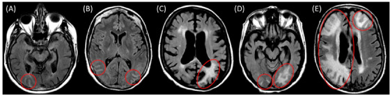
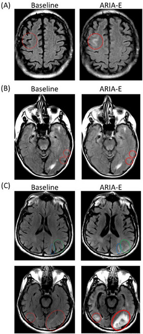
Introduction: Anti-amyloid-β (Aβ) monoclonal antibodies (mAbs) offer the promise of disease modification and are emerging treatment options in Alzheimer's disease. Anti-Aβ mAbs require brain magnetic resonance imaging (MRI) examinations to detect anti-amyloid-induced amyloid-related imaging abnormalities (ARIA), important adverse drug reactions associated with some anti-Aβ mAbs currently available in the United States and in clinical development. We present a simple rating system for ARIA-edema (ARIA-E) that can assess severity on a 3- or 5-point scale based upon a single linear measurement of the largest area of lesion, and dissemination in space, termed the 3-point Severity Scale of ARIA-E (SSAE-3) and the 5-point Severity Scale of ARIA-E (SSAE-5), respectively.
Methods: MRI results were collected from 75 participants from the SCarlet RoAD (NCT01224106) and Marguerite RoAD (NCT02051608) studies of gantenerumab. Three neuroradiologists experienced with the detection of ARIA-E were selected to read all cases independently. One rater was then chosen for a second read to assess intra-reader reproducibility.
Results: The three raters had high agreement in identifying and grading ARIA-E. The Cohen/Fleiss kappa (κ) scores (95% confidence interval [CI]) for the inter- and intra-reader comparisons for SSAE-3 and SSAE-5 were 0.79 (0.70-1.00), 0.94 (0.94-1.00), 0.73 (0.66-1.00), and 0.90 (0.90-1.00), respectively.
Discussion: Our study suggests that SSAE-3 and SSAE-5 are valid ARIA-E rating scales for use in routine clinical practice by experienced radiologists in specialized settings. The application of these scales in everyday use in clinical practice will support the expansion of anti-Aβ mAbs as a treatment option for people living with Alzheimer's disease.
Highlights: A simple rating scale is needed to rate severity of amyloid-related imaging abnormalities-edema (ARIA-E) in both research and clinical settings.The 3- and 5-point Severity Scales of ARIA-E (SSAE-3/-5) have good inter- and intra-reader agreement.The SSAE-3/-5 have been used in most major Alzheimer's disease (AD) trials to date and are suitable for large-scale use in routine clinical practice, which may help support the expansion of anti-amyloid antibodies as treatment options for AD.
Keywords: Alzheimer's disease; amyloid; amyloid beta; amyloid plaques; diagnostic imaging.
© 2023 The Authors. Alzheimer's & Dementia: Diagnosis, Assessment & Disease Monitoring published by Wiley Periodicals LLC on behalf of Alzheimer's Association.
Conflict of interest statement
Luc Bracoud is a full‐time employee of Clario, Inc. (formerly known as Bioclinica, Inc.). Gregory Klein is a full‐time employee of and shareholder in F. Hoffmann‐La Roche Ltd. Marco Lyons is a full‐time employee of and shareholder in Roche Products Ltd. Marzia A. Scelsi is a full‐time employee of Roche Products Ltd. Jakub Wojtowicz is a full‐time employee of and shareholder in F. Hoffmann‐La Roche Ltd. Szofia Bullain is a full‐time employee of and shareholder in F. Hoffmann‐La Roche Ltd. Derk Purcell reports outside the submitted work personal fees from Biogen and provides both consultative services and image interpretation for Clario, Inc. Jochen B. Fiebach reports outside the submitted work personal fees from AbbVie, AC Immune, Artemida, Bioclinica/Clario, Biogen, BMS, Brainomix, Cerevast, Daiichi Sankyo, Eisai, F. Hoffmann‐La Roche AG, Eli Lilly, Guerbet, Ionis Pharmaceuticals, IQVIA, Janssen, Julius Clinical, jung diagnostics, Lysogene, Merck, Nicolab, Premier Research, and TauRx. Jerome Barakos provides both consultative services and image interpretation for Clario, Inc. Joyce Suhy is a full‐time employee of Clario, Inc.
Figures
References
-
- Hardy J, Selkoe DJ, The amyloid hypothesis of Alzheimer's disease: progress and problems on the road to therapeutics. Science. 2002;297(5580):353‐356. - PubMed
-
- Villemagne VL, Burnham S, Bourgeat P, et al. Amyloid β deposition, neurodegeneration, and cognitive decline in sporadic Alzheimer's disease: a prospective cohort study. Lancet Neurol. 2013;12(4):357‐367. - PubMed
-
- Thal DR, Walter J, Saido TC, et al. Neuropathology and biochemistry of Aβ and its aggregates in Alzheimer's disease. Acta Neuropathol. 2015;129(2):167‐182. - PubMed
LinkOut - more resources
Full Text Sources