

Morphologic alterations of the fear circuitry: the role of sex hormones and oral contraceptives
- PMID: 38027091
- PMCID: PMC10661904
- DOI: 10.3389/fendo.2023.1228504
Morphologic alterations of the fear circuitry: the role of sex hormones and oral contraceptives
Abstract
Background: Endogenous sex hormones and oral contraceptives (OCs) have been shown to influence key regions implicated in fear processing. While OC use has been found to impact brain morphology, methodological challenges remain to be addressed, such as avoiding selection bias between OC users and non-users, as well as examining potential lasting effects of OC intake.
Objective: We investigated the current and lasting effects of OC use, as well as the interplay between the current hormonal milieu and history of hormonal contraception use on structural correlates of the fear circuitry. We also examined the role of endogenous and exogenous sex hormones within this network.
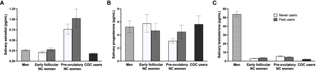
Methods: We recruited healthy adults aged 23-35 who identified as women currently using (n = 62) or having used (n = 37) solely combined OCs, women who never used any hormonal contraceptives (n = 40), or men (n = 41). Salivary endogenous sex hormones and current users' salivary ethinyl estradiol (EE) were assessed using liquid chromatography - tandem mass spectrometry. Using structural magnetic resonance imaging, we extracted surface-based gray matter volumes (GMVs) and cortical thickness (CT) for regions of interest of the fear circuitry. Exploratory whole-brain analyses were conducted with surface-based and voxel-based morphometry methods.
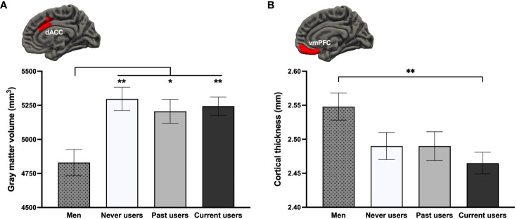
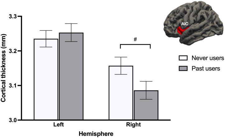
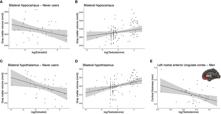
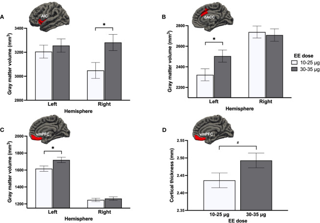
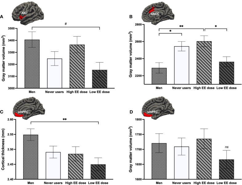
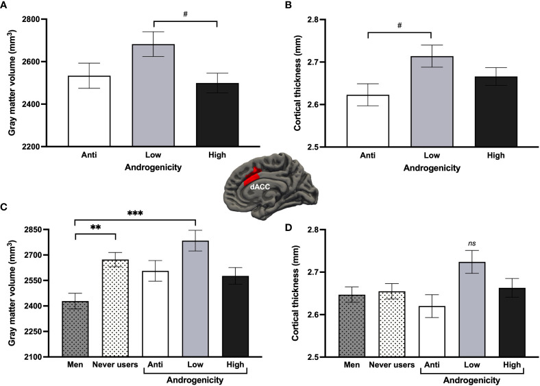
Results: Compared to men, all three groups of women exhibited a larger GMV of the dorsal anterior cingulate cortex, while only current users showed a thinner ventromedial prefrontal cortex. Irrespective of the menstrual cycle phase, never users exhibited a thicker right anterior insular cortex than past users. While associations with endogenous sex hormones remain unclear, we showed that EE dosage in current users had a greater influence on brain anatomy compared to salivary EE levels and progestin androgenicity, with lower doses being associated with smaller cortical GMVs.
Discussion: Our results highlight a sex difference for the dorsal anterior cingulate cortex GMV (a fear-promoting region), as well as a reduced CT of the ventromedial prefrontal cortex (a fear-inhibiting region) specific to current OC use. Precisely, this finding was driven by lower EE doses. These findings may represent structural vulnerabilities to anxiety and stress-related disorders. We showed little evidence of durable anatomical effects, suggesting that OC intake can (reversibly) affect fear-related brain morphology.
Keywords: cortical thickness; fear circuitry; gray matter volume; oral contraceptives; sex hormones; structural MRI.
Copyright © 2023 Brouillard, Davignon, Turcotte and Marin.
Conflict of interest statement
The authors declare that the research was conducted in the absence of any commercial or financial relationships that could be construed as a potential conflict of interest.
Figures
Similar articles
-
Starting the pill during adolescence: Age of onset and duration of use influence morphology of the hippocampus and ventromedial prefrontal cortex.Eur J Neurosci. 2024 Oct;60(8):5876-5899. doi: 10.1111/ejn.16509. Epub 2024 Sep 8. Eur J Neurosci. 2024. PMID: 39245916
-
Comparative contraceptive efficacy and mechanism of action of the norgestimate-containing triphasic oral contraceptive.Acta Obstet Gynecol Scand Suppl. 1992;156:9-14. doi: 10.3109/00016349209156509. Acta Obstet Gynecol Scand Suppl. 1992. PMID: 1324557 Clinical Trial.
-
Effect of two oral contraceptives containing ethinylestradiol and gestodene or norgestimate upon androgen parameters and serum binding proteins.Contraception. 1995 Jun;51(6):341-6. doi: 10.1016/0010-7824(95)00098-u. Contraception. 1995. PMID: 7554973 Clinical Trial.
-
[Low dose oral contraceptives: 30 microg are still used?].Minerva Ginecol. 2009 Oct;61(5):453-8. Minerva Ginecol. 2009. PMID: 19749677 Review. Italian.
-
[Individualization of low-dose oral contraceptives. Pharmacological principles and practical indications for oral contraceptives].Minerva Ginecol. 2007 Aug;59(4):415-25. Minerva Ginecol. 2007. PMID: 17923832 Review. Italian.
Cited by
-
Breaking New Ground With Endoxifen: Augmentation Strategies in OCD Management-A Case Series.Case Rep Psychiatry. 2025 Mar 26;2025:2908673. doi: 10.1155/crps/2908673. eCollection 2025. Case Rep Psychiatry. 2025. PMID: 40177005 Free PMC article.
-
Subjective, behavioural and physiological correlates of stress in women using hormonal contraceptives.Br J Psychiatry. 2025 Jun;226(6):392-400. doi: 10.1192/bjp.2025.7. Epub 2025 Jun 13. Br J Psychiatry. 2025. PMID: 40511505 Free PMC article.
-
Current oral contraceptive use affects explicit and implicit measures of depression in women.Front Psychol. 2024 Oct 18;15:1462891. doi: 10.3389/fpsyg.2024.1462891. eCollection 2024. Front Psychol. 2024. PMID: 39492815 Free PMC article.
References
-
- Ekman P. An argument for basic emotions. Cogn Emotion (1992) 6(3/4):169–200. doi: 10.1080/02699939208411068 - DOI
MeSH terms
Substances
LinkOut - more resources
Full Text Sources
Miscellaneous