. 2023 Nov 4;9(11):e21850.
doi: 10.1016/j.heliyon.2023.e21850. eCollection 2023 Nov.

β-Cyclodextrin capped ZnS nanoparticles for CER-assisted colorimetric and spectrophotometric detection of Pb2⁺, Cu2⁺, and Hg2⁺ in an aqueous solution

Affiliations

β-Cyclodextrin capped ZnS nanoparticles for CER-assisted colorimetric and spectrophotometric detection of Pb2⁺, Cu2⁺, and Hg2⁺ in an aqueous solution

Vyshnavi T Veetil et al. Heliyon. .

Abstract

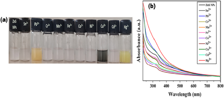
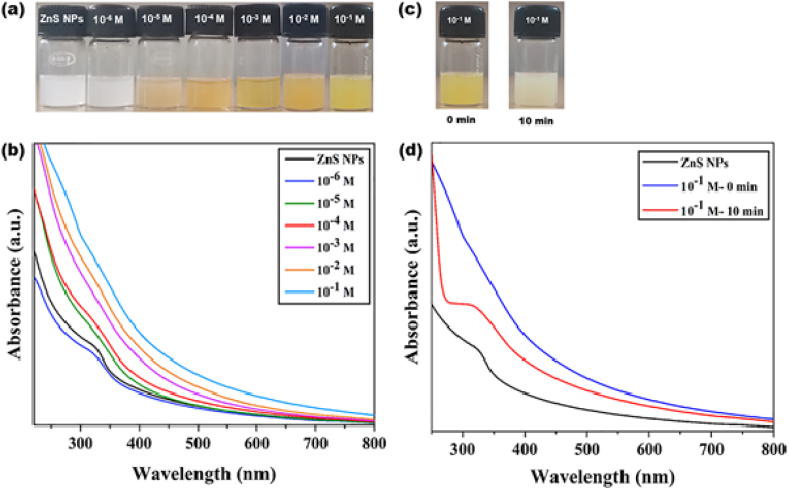
Herein, simple, low-cost, and room-temperature synthesis of beta-cyclodextrin (β-CD) stabilized zinc sulfide nanoparticle (ZnS NP) through the chemical precipitation method has been reported for cation exchange reaction (CER) based colorimetric sensing of Pb2+, Cu2+, and Hg2+. Formation of β-CD stabilized ZnS NPs (ZnS@β-CD) was verified by physicochemical characterization techniques such as XRD, XPS, FE-SEM, and TEM. ZnS@β-CD NPs showed color change selectively for the metal ions Pb2⁺, Cu2⁺, and Hg2⁺ among the various metal ions including Sn2⁺, Cr³⁺, Mn2⁺, Fe³⁺, Co2⁺, Ni2⁺, and Cd2⁺. The solubility product of reactants and the transformed products are the reason for selective CER of ZnS@β-CD NPs towards Pb2⁺, Cu2⁺, and Hg2⁺ ions. ZnS@β-CD NPs dispersion revealed rapid color change from white to orange, black, and bright yellow on the addition of higher concentrations of Pb2⁺, Cu2⁺, and Hg2⁺ respectively. This color change is due to the formation of complete CER-transformed nanostructures such as PbS, CuS, and HgS in higher concentrations (10⁻1- 10⁻³ M) of corresponding metal ions. The partial CER altered products Zn1-x,PbxS, Zn1-xCuxS and Zn1-xHgxS were detected due to the appearance of pale color in the lower metal ions concentrations of 10⁻⁴ - 10⁻⁶ M. This CER assisted transformation was also monitored through spectrophotometric methods. Moreover, infrared spectroscopic analysis was used to testify the structure of CER transformed product. The synthesized ZnS@β-CD NPs act as an efficient CER-based sensor for distinguishing and determining Pb2⁺, Cu2⁺, and Hg2⁺ at different level concentrations in the aqueous solution.

Keywords: Cation exchange reactions; Colorimetric sensor; Heavy metal ions; ZnS nanoparticles; β-Cyclodextrin.


Conflict of interest statement

The authors declare that they have no known competing financial interests or personal relationships that could have appeared to influence the work reported in this paper.

Figures

Image 1
Graphical abstract
Scheme 1
Scheme 1
Schematic diagram for the synthesis of ZnS@β-CD NPs.
Fig. 1
Fig. 1
XRD patterns of ZnS@β-CD NPs.
Fig. 2
Fig. 2
The XPS analysis of ZnS@β-CD NPs (a) Wide scan XPS (b) Zn 2p deconvoluted peaks (c) S 2p deconvoluted peaks (d) C 1s deconvoluted peaks, (e) O 1s deconvoluted peaks.
Fig. 3
Fig. 3
(a) SEM Image of β-CD, (b) SEM image of ZnS@β-CD NPs, (c) EDX spectra of ZnS@β-CD NPs, (d) Elemental mapping images of ZnS@β-CD NPs.
Fig. 4
Fig. 4
HR-TEM images of ZnS@β-CD NPs at various scale (a) 50 nm, (b) 10 nm, (c) Particle size distribution histogram, (d) SAED pattern.
Fig. 5
Fig. 5
(a) UV–Visible absorption spectrum and (b) Tauc plot of ZnS@β-CD NPs.
Fig. 6
Fig. 6
(a) Colorimetric and (b) UV–Visible spectroscopic analysis of ZnS@β-CD NPs towards different metal ions.
Fig. 7
Fig. 7
(a) Colorimetric and (b) UV–Visible spectroscopic analysis of ZnS NPs (without β-CD) towards different metal ions.
Fig. 8
Fig. 8
Colorimetric responses of ZnS@β-CD NPs towards Pb2⁺, Cu2⁺, and Hg2⁺ in presence of other heavy metal ions.
Fig. 9
Fig. 9
(a) Colorimetric and (b) UV–Visible spectroscopic analysis of ZnS@β-CD NPs towards different concentrations of Pb2⁺. Time-resolved interactions of ZnS@β-CD NPs with Pb2⁺: (c) Photographs of 10⁻2 M and 10⁻1 M of Pb2⁺ at 0 min and after time t (50 min for 10⁻2 M and 20 min for 10⁻1 M). (d) Time-based UV–Visible spectroscopic analysis of 10⁻2 M and 10⁻1 M of Pb2⁺ metal ions.
Fig. 10
Fig. 10
(a) Colorimetric and (b) UV–Visible spectroscopic measurements of ZnS@β-CD NPs towards different concentrations of Cu2⁺, (c) XRD pattern of CuS NS.
Fig. 11
Fig. 11
(a) Colorimetric and (b) UV–Visible spectroscopic measurements of ZnS@β-CD NPs towards different concentrations of Hg2⁺. Time-resolved interactions of ZnS@β-CD NPs with Hg2+: (c) Photograph of 10⁻1 M of Hg2⁺ at 0 min and after 10 min. (d) Time-based UV–Visible spectroscopic responses of 10⁻1 M of Hg2⁺.
Fig. 12
Fig. 12
(a) Colorimetric and (b) UV–Visible spectroscopic measurements of ZnS@β-CD NP added with multiple metal ions (10⁻³ M of Pb2⁺, Cu2⁺, &Hg2⁺, Pb2⁺ & Hg2⁺, Cu2⁺ & Hg2⁺, and Pb2⁺ & Cu2⁺) at 0 min and 10 min.
Fig. 13
Fig. 13
Colorimetric responses of ZnS@β-CD towards 10⁻³ M concentrations of Pb2⁺, Cu2⁺, and Hg2⁺ metal ions (a) at 40 °C and (b) at 50 °C. UV–Visible spectroscopic measurements of ZnS@β-CD NPs towards 10⁻³ M concentrations of Pb2⁺, Cu2⁺, and Hg2⁺ metal ions (c) at 40 °C and (d) at 50 °C.
Fig. 14
Fig. 14
(a) Colorimetric and (b–g) UV–Visible spectroscopic measurements of ZnS@β-CD NP in different pH medium in presence of 10⁻³ M concentrations of Pb2⁺, Cu2⁺ and Hg2⁺.
Fig. 15
Fig. 15
FT-IR spectra of ZnS@β-CD NPs and its CER converted products.

References

    1. Kalarikkandy A.V., Sree N., Ravichandran S., Dheenadayalan G. Copolymer - MnO2 nanocomposites for the adsorptive removal of organic pollutants from water. Environ. Sci. Pollut. Control Ser. 2023;30:71454–71463. doi: 10.1007/s11356-022-22137-2. - DOI - PubMed
    1. A.V.K K., Gangadharan D. Nano-Structures & Nano-Objects Adsorptive remediation of organic pollutant and arsenic (V) ions from water using Fe3O4 -MnO2 nanocomposite. Nano-Structures & Nano-Objects. 2022;29 doi: 10.1016/j.nanoso.2022.100837. - DOI
    1. Jaishankar M., Tseten T., Anbalagan N., Mathew B.B., Beeregowda K.N. Toxicity, mechanism and health effects of some heavy metals. Interdiscipl. Toxicol. 2014;7:60–72. doi: 10.2478/intox-2014-0009. - DOI - PMC - PubMed
    1. Tchounwou P.B., Yedjou C.G., Patlolla A.K., Sutton D.J. Heavy metal toxicity and the environment. Molecular, Clinical and Environ. Toxicicol. 2012;3:133–164. doi: 10.1007/978-3-7643-8340-4. - DOI - PMC - PubMed
    1. Aoki K., Aschner M., Cullen J.T., Farkas E., Filella M., Hauser P., Klotz K., Küpper H., Maret W., Stewart T.J., Stillman M.J., Pecoraro V.L., Pohl H.R., Tylkowski B. Walter de Gruyter GmbH & Co KG; 2017. Lead: its Effects on Environment and Health; p. 17. - DOI

LinkOut - more resources