Review
. 2023 Nov 16;19(5):26-36.
doi: 10.14797/mdcvj.1309. eCollection 2023.

Metabolic Control of Cardiomyocyte Cell Cycle

Affiliations
Review

Metabolic Control of Cardiomyocyte Cell Cycle

Ivan Menendez-Montes et al. Methodist Debakey Cardiovasc J. .

Abstract

Current therapies for heart failure aim to prevent the deleterious remodeling that occurs after MI injury, but currently no therapies are available to replace lost cardiomyocytes. Several organisms now being studied are capable of regenerating their myocardium by the proliferation of existing cardiomyocytes. In this review, we summarize the main metabolic pathways of the mammalian heart and how modulation of these metabolic pathways through genetic and pharmacological approaches influences cardiomyocyte proliferation and heart regeneration.

Keywords: cardiac regeneration; cardiomyocyte proliferation; glycolysis; oxidative phosphorylation; reactive oxygen species; uridine diphosphate N-acetylglucosamine (UDP GlycNAC).


Conflict of interest statement

HAS is supported by NIH R01 HL149137-01, NIH 1P01HL160476-01A1, NIH R35 HL166563-01, and NIH P01HL160488. IM-M is supported by AHA Postdoctoral Fellowship 903385.The authors have no competing interests to declare.

Figures

Cardiac metabolism from embryo to adult. Schematic representation of glycolysis, glucose oxidation, and fatty acid oxidation during embryonic, neonatal, and adult stages. The graph also includes the temporal dynamic of cardiac key processes (proliferation, binucleation, and DNA damage) in relation to the metabolic changes
Figure 1
Cardiac metabolism from embryo to adult. Schematic representation of glycolysis, glucose oxidation, and fatty acid oxidation during embryonic, neonatal, and adult stages. The graph also includes the temporal dynamic of cardiac key processes (proliferation, binucleation, and DNA damage) in relation to the metabolic changes.
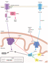
Glucose and fatty acids metabolism in the adult heart. Glucose oxidation (purple), fatty acids metabolism (blue), and Pentose Phosphate Pathway (pink) representation. Genetic/pharmacological models are indicated for activation/overexpression (green + symbol) or inhibition/deletion (red cross). Their outcomes are indicated on the right side of the name for increased (green up arrow) or decreased (red down arrow) proliferation/regeneration. Signaling pathways are indicated with a yellow star
Figure 2
Glucose and fatty acids metabolism in the adult heart. Glucose oxidation (purple), fatty acids metabolism (blue), and Pentose Phosphate Pathway (pink) representation. Genetic/pharmacological models are indicated for activation/overexpression (green + symbol) or inhibition/deletion (red cross). Their outcomes are indicated on the right side of the name for increased (green up arrow) or decreased (red down arrow) proliferation/regeneration. Signaling pathways are indicated with a yellow star.
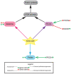
Contributions of amino acid metabolism in the adult heart. Contributions of glutamine (pink), branched-chain amino acids (BCAAs, purple) and proline (blue) to cardiac regeneration. Genetic/pharmacological models are indicated for activation/overexpression (green + symbol) or inhibition/deletion (red cross). Their outcomes are indicated on the right side of the name for increased (green up arrow) or decreased (red down arrow) proliferation/regeneration
Figure 3
Contributions of amino acid metabolism in the adult heart. Contributions of glutamine (pink), branched-chain amino acids (BCAAs, purple) and proline (blue) to cardiac regeneration. Genetic/pharmacological models are indicated for activation/overexpression (green + symbol) or inhibition/deletion (red cross). Their outcomes are indicated on the right side of the name for increased (green up arrow) or decreased (red down arrow) proliferation/regeneration.

References

    1. Porrello ER, Mahmoud AI, Simpson E, et al. Transient regenerative potential of the neonatal mouse heart. Science. 2011. Feb 25;331(6020):1078-80. doi: 10.1126/science.1200708 - DOI - PMC - PubMed
    1. Soonpaa MH, Kim KK, Pajak L, Franklin M, Field LJ. Cardiomyocyte DNA synthesis and binucleation during murine development. Am J Physiol. 1996. Nov;271(5 Pt 2):H2183-9. doi: 10.1152/ajpheart.1996.271.5.H2183 - DOI - PubMed
    1. Lopaschuk GD, Jaswal JS. Energy metabolic phenotype of the cardiomyocyte during development, differentiation, and postnatal maturation. J Cardiovasc Pharmacol. 2010. Aug;56(2):130-40. doi: 10.1097/FJC.0b013e3181e74a14 - DOI - PubMed
    1. Makinde AO, Kantor PF, Lopaschuk GD. Maturation of fatty acid and carbohydrate metabolism in the newborn heart. Mol Cell Biochem. 1998. Nov;188(1-2):49-56. - PubMed
    1. Puente BN, Kimura W, Muralidhar SA, et al. The oxygen-rich postnatal environment induces cardiomyocyte cell-cycle arrest through DNA damage response. Cell. 2014. Apr 24;157(3):565-79. doi: 10.1016/j.cell.2014.03.032. Erratum in: Cell. 2014 May 22;157(5):1243 - DOI - PMC - PubMed