. 2023 Nov 29;14(1):7864.
doi: 10.1038/s41467-023-43403-y.

Illuminating the mechanism and allosteric behavior of NanoLuc luciferase

Affiliations

Illuminating the mechanism and allosteric behavior of NanoLuc luciferase

Michal Nemergut et al. Nat Commun. .

Abstract

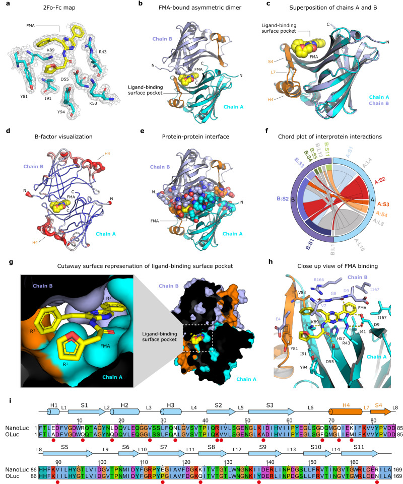
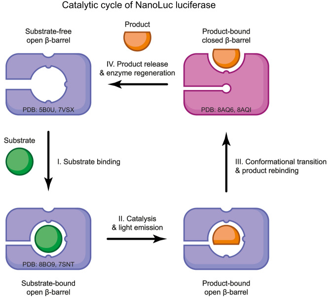
NanoLuc, a superior β-barrel fold luciferase, was engineered 10 years ago but the nature of its catalysis remains puzzling. Here experimental and computational techniques are combined, revealing that imidazopyrazinone luciferins bind to an intra-barrel catalytic site but also to an allosteric site shaped on the enzyme surface. Structurally, binding to the allosteric site prevents simultaneous binding to the catalytic site, and vice versa, through concerted conformational changes. We demonstrate that restructuration of the allosteric site can boost the luminescent reaction in the remote active site. Mechanistically, an intra-barrel arginine coordinates the imidazopyrazinone component of luciferin, which reacts with O2 via a radical charge-transfer mechanism, and then it also protonates the resulting excited amide product to form a light-emitting neutral species. Concomitantly, an aspartate, supported by two tyrosines, fine-tunes the blue color emitter to secure a high emission intensity. This information is critical to engineering the next-generation of ultrasensitive bioluminescent reporters.


Conflict of interest statement

The authors declare no competing interests.

Figures

Fig. 1
Fig. 1. The structure of FMA bound in the ligand-binding surface pocket of NanoLuc luciferase.
a 2Fo-Fc electron density (contour level 1.2 σ) at the FMA-oxyluciferin binding site. b Cartoon representation of the overall structure of NanoLuc asymmetric dimer (chain A in cyan and chain B in blue) with bound FMA luciferin (yellow). c Superposition of chain A (cyan) and chain B (blue). The structural element responsible for symmetry breaking, encompassing helix H4, loop L7 and strand S4, is colored orange. d B-factor putty representation of NanoLuc homodimer. e Dimer interface visualization. All residues involved in the dimer interface are shown as space-filling spheres. FMA is shown as yellow spheres. f Chord plot showing the interactions between chains A and B in the NanoLuc dimer at the secondary structure level, calculated and visualized by Protein Contact Atlas. g Cutaway surface representation of FMA-bound NanoLuc dimer. h Close-up view of FMA-binding pocket with residues creating the active site in stick representation. Key hydrogen bonds are shown as dashed yellow lines. i Sequence alignment between NanoLuc and the catalytic unit of O. gracilirostris luciferase (OLuc). Secondary structure elements found in NanoLuc are shown above the alignment. Amino acid residues mutated during the NanoLuc engineering are labeled with the red dot. The numbering indicated above the alignment corresponds to the NanoLuc structure (PDB ID: 5B0U).
Fig. 2
Fig. 2. The structure of CEI-bound crystallographic homotetramer of NanoLuc luciferase.
a 2Fo-Fc electron density (contour level 1.2 σ) at the CEI-oxyluciferin binding site. b The overall structure of the NanoLuc homotetramer, composed of two tail-to-tail asymmetric homodimers (chain A in cyan, chain B in blue, chain C in violet and chain D in green). The CEI luciferin is shown as space-filling spheres (green). c Cutaway surface representation of CEI-bound NanoLuc tetramer. d Close-up views of CEI-binding pocket with residues creating the active site in stick representation. Key hydrogen bonds are shown as dashed yellow lines. e Superposition of FMA (yellow) and CEI (green) binding modes.
Fig. 3
Fig. 3. Conformational switch between open and closed NanoLuc β-barrel structures.
a Cartoon representations of (i) ligand-free apo-NanoLuc structure (PDB ID: 5B0U), (iii) FMA-bound NanoLuc structure, and (ii) their superposition. The H93 and Y94 residues and FMA-luciferin are shown as space-filling spheres. b Close-up views of (i) luciferin-binding surface pocket in ligand-free apo-NanoLuc structure (PDB ID: 5B0U), (iii) FMA-bound NanoLuc structure, and (ii) their superposition. R43, H57, K89, H93 and Y94 residues are shown as space-filling spheres, and the FMA luciferin is shown as yellow sticks. c 2Fo-Fc electron density (contour level 1.2 σ) at the chloride-binding sites 1 and 2. d, e Cutaway surface representations of NanoLuc β-barrel interior in a closed (d) and open (e) state. Note that in the closed β-barrel state, the two chloride ions are bound inside the β-barrel.
Fig. 4
Fig. 4. Characterization of NanoLuc mutants.
a Heatmap showing the relative luciferase activities of NanoLuc mutants with CTZ (left column) and FMZ (right column) luciferins. NanoLuc wild-type = 100%. b Structures of FMA-bound asymmetric dimer of NanoLuc wild-type (i) and luciferin-free symmetric dimer of NanoLuc-Y94A mutant (ii), and their superposition (iii). The tyrosine-tyrosine gate Y81 and Y94 (A94) residues and FMA-luciferin are shown as space-filling spheres. c Close-up view of the CEI-bound luciferin-binding surface site of NanoLuc luciferase. Three residues, namely D9, H57 and K89, whose mutations increase bioluminescence with CTZ are shown as cyan space-filling spheres. The other protein residues are shown as cyan sticks, and the CEI-luciferin is shown as green sticks. d Heatmap showing the relative luciferase activities of NanoLuc mutants with CTZ (left column) and FMZ (right column) luciferins; NanoLuc wild-type = 100%. e Relative fold increases ± standard errors (s.e.) of kinetic parameters (Km, kcat and kcat/Km) observed in the double NanoLuc-D9R/K89R and triple NanoLuc-D9R/H57A/K89R mutants; NanoLuc wild-type = 1. Absolute values of kinetic parameters (Km, kcat and kcat/Km) are summarized in Table 1. f, g Long-term live-cell bioluminescence imaging of cultured ARPE-19 cells expressing either original NanoLuc or engineered NanoLucCTZ and powered by EnduRen (f) or Nano-Glo Endurazine (g) substrate. The luciferase activities upon addition of the corresponding luciferin were measured by LuminoCell device, integration time was 5 min. Each data point represents the mean enzymatic activity of three replicates. The standard error is represented as a shaded area around the mean curve.
Fig. 5
Fig. 5. Catalytic mechanism for NanoLuc-type luciferase reaction.
a 2Fo-Fc electron density map (contour level 3.0 σ) of azaCTZ bound in the intra-barrel catalytic site of NanoLucCTZ. b Cartoon representation of the overall structure of NanoLucCTZ (cyan) with bound azaCTZ (green). c Close-up view of azaCTZ (green) bound to NanoLucCTZ, with important residues creating the active site in cyan stick representation. Red sphere; a water molecule bound in the catalytic site. Key molecular contacts are shown as dashed yellow lines. d Mutagenesis of NanoLuc active site residues. Data indicate the average relative luciferase activities of each mutant. Assays were done in triplicate; error bars represent standard deviations. e Visualizations of the NanoLucCTZ catalytic site with modeled CTZ (i), 2-peroxy-CTZ (ii), CTZ dioxetanone (iii), and CEI (iv). Key protein residues are shown as sticks and lines, molecular oxygen (OXY) is shown in red sphere, and hydrogen bonds are shown as dashed lines. f A blueprint for NanoLuc-type reaction mechanism. The cycle starts with the binding of CTZ into the catalytic site localized inside the β-barrel structure, and it enters with a deprotonated imidazopyrazinone core, as demonstrated experimentally in previous work. I. Upon the entry, the -OH group of the C6-(p-hydroxyphenyl) moiety is hydrogen-bonded with two tyrosines (Y94 and Y114), as well as deprotonated by D139, yielding the activated dianion O10-CTZ. Then, the side chain of R162, and perhaps helped by the side chain of Q42, position a co-substrate molecule (dioxygen) such that it can be attacked by the C2 carbon of O10-CTZ. II. The initial interaction proceeds via a charge-transfer radical mechanism. III. The next steps encompasses radical pairing and termination, resulting in the 2-peroxy-CTZ anion, which then undergoes intramolecular cyclization via a nucleophilic addition-elimination mechanism. IV. This cyclization generates a dioxetanone intermediate with a deprotonated amide group. V. The side chain of R162 protonates the amide group of oxyluciferin in order to avoid attenuation of the luminescence. VI. The energy-rich dioxetanone intermediate is unstable and decomposes by decarboxylation into an excited CEI product. VII. When returning to the ground state, the excited CEI releases a blue photon. Finally, the protonation status of R162 is restored by the proton transfer from a water molecule.
Fig. 6
Fig. 6. Molecular docking and computational simulations of NanoLuc complexes.
a Visualization of the best poses of FMZ (magenta sticks) from molecular docking to the NanoLuc monomer dimer (chain A cyan and chain B blue cartoon), in comparison with the crystal-bound FMA (yellow sticks). b Two potential access tunnels (yellow and blue spheres) to the central luciferin binding cavity (green spheres) of NanoLucCTZ (salmon cartoon) are shown from the front and the side view. The tunnels were computed on Caver Web. c The final poses of CTZ (orange sticks) from ASMD simulations of NanoLuc structures in the closed and open β-barrel state, and NanoLucCTZ open β-barrel structure. The crystal-bound azaCTZ (transparent green sticks) is shown in each picture for reference. The interacting aromatic residues are shown as thin sticks. The distances (in Å) between H2/H3 and H3/H4 helices are indicated by yellow dashed lines. Associated MD trajectories are shown in Supplementary Movies 1–3. d NanoLuc closed β-barrel structure with shown mutations (L27V, K33N, K43R, and Y68D) introduced during the last third phase of NanoLuc development responsible for maximizing FMZ fluorescence. e Equilibrium probabilities of FMZ macrostates bound in the surface pocket of monomeric and dimeric NanoLuc. The calculation was bootstrapped 100 times using a random 80% of the data. The height of the bars corresponds to the mean value and the error bars show the standard deviation. f The potential of mean force needed to steer the two subunits of different NanoLuc dimers up to 12 Å apart, obtained by adaptive steered molecular dynamics.
Fig. 7
Fig. 7. Structure-based model for NanoLuc luciferase action.
I. A substrate molecule enters the intra-barrel catalytic site, the luciferase retains an “open β-barrel” conformation. II. Catalytic conversion of the substrate into its reaction product, followed by the emission of a blue photon. III. Release of the product out of the intra-barrel catalytic site. This step is accompanied by an open-to-closed conformational transition and a rebinding of the product to the newly formed allosteric site on the protein surface. IV. Dissociation of the product from the surface allosteric site, allowing recycling into the pre-catalytic “open β-barrel” state. The PDB ID codes for representative crystallographic structures are provided.

References

    1. Haddock SHD, Moline MA, Case JF. Bioluminescence in the sea. Ann. Rev. Mar. Sci. 2010;2:443–493. doi: 10.1146/annurev-marine-120308-081028. - DOI - PubMed
    1. Mitiouchkina T, et al. Plants with genetically encoded autoluminescence. Nat. Biotechnol. 2020;38:944–946. doi: 10.1038/s41587-020-0500-9. - DOI - PMC - PubMed
    1. Schenkmayerova A, et al. Engineering the protein dynamics of an ancestral luciferase. Nat. Commun. 2021;12:3616. doi: 10.1038/s41467-021-23450-z. - DOI - PMC - PubMed
    1. Su Y, et al. Novel NanoLuc substrates enable bright two-population bioluminescence imaging in animals. Nat. Methods. 2020;17:852–860. doi: 10.1038/s41592-020-0889-6. - DOI - PMC - PubMed
    1. Syed AJ, Anderson JC. Applications of bioluminescence in biotechnology and beyond. Chem. Soc. Rev. 2021;50:5668–5705. doi: 10.1039/D0CS01492C. - DOI - PubMed

Publication types