. 2023 Nov 29;23(1):274.
doi: 10.1186/s12911-023-02362-6.

Automatic deep learning-based pleural effusion segmentation in lung ultrasound images

Affiliations

Automatic deep learning-based pleural effusion segmentation in lung ultrasound images

Damjan Vukovic et al. BMC Med Inform Decis Mak. .

Abstract

Background: Point-of-care lung ultrasound (LUS) allows real-time patient scanning to help diagnose pleural effusion (PE) and plan further investigation and treatment. LUS typically requires training and experience from the clinician to accurately interpret the images. To address this limitation, we previously demonstrated a deep-learning model capable of detecting the presence of PE on LUS at an accuracy greater than 90%, when compared to an experienced LUS operator.

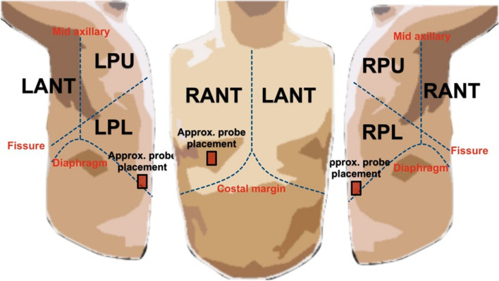
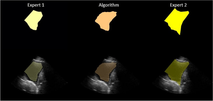
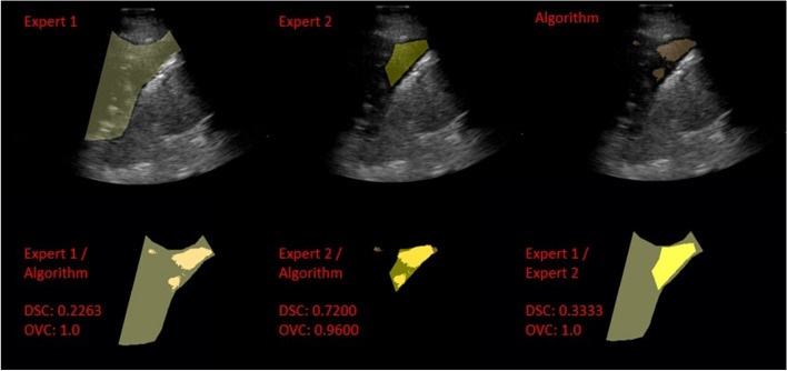
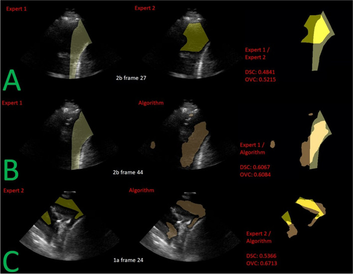
Methods: This follow-up study aimed to develop a deep-learning model to provide segmentations for PE in LUS. Three thousand and forty-one LUS images from twenty-four patients diagnosed with PE were selected for this study. Two LUS experts provided the ground truth for training by reviewing and segmenting the images. The algorithm was then trained using ten-fold cross-validation. Once training was completed, the algorithm segmented a separate subset of patients.

Results: Comparing the segmentations, we demonstrated an average Dice Similarity Coefficient (DSC) of 0.70 between the algorithm and experts. In contrast, an average DSC of 0.61 was observed between the experts.

Conclusion: In summary, we showed that the trained algorithm achieved a comparable average DSC at PE segmentation. This represents a promising step toward developing a computational tool for accurately augmenting PE diagnosis and treatment.

Keywords: Deep Learning; Lung Ultrasound; Pleural Effusion / diagnostic imaging; Point-of-Care Ultrasound.


Conflict of interest statement

The authors declare no competing interests.

Figures

Fig. 1
Fig. 1
Examples of the scanning regions (viz. Right Anterior (RANT), Right Posterior Lower (RPL), Left Posterior Lower (LPL)) and the approximate probe placement during the image acquisition of LUS frames containing PE. Figure created and owned by coauthors
Fig. 2
Fig. 2
A flow diagram showing the pathology distribution of the original PE patients to the final training/validation/test dataset split used to train the algorithm
Fig. 3
Fig. 3
Comparison of PE identified LUS frames, where image (A) does not meet the clinical criteria needed for contouring PE and image (B) is the ideal case for PE segmentation. The presence of PE was defined as an anechoic space (in red) between the parietal pleura (lining the chest wall in orange and diaphragm in yellow) and visceral pleura (lining the lung surface in green) image B, along with the collapsed lung tissue (in green)
Fig. 4
Fig. 4
An example of a LUS image and corresponding segmentations generated by expert 1, expert 2, and the algorithm (from left to right) respectively. On the bottom row, the LUS image and the segmentation are overlayed; on the top row, only segmentations are shown
Fig. 5
Fig. 5
An example of a LUS image and corresponding segmentations generated by expert 1, expert 2 and the algorithm (from left to right), respectively, extracted from the worst performing video in terms of DSC (i.e., 4b from Table 8). Overlays of LUS image and segmentation are shown in the top row. The bottom row shows the DSC and OVC scores calculated between algorithm / expert 1, algorithm / expert 2, and expert 1 / expert 2 segmentations
Fig. 6
Fig. 6
An example of the corresponding segmentations from expert 1, expert 2 and the algorithm’s prediction shown overlayed over the LUS frame of the worst performing DSC video (i.e., 2b and 1a from Table 11). Each row (A, B, C) shows the worst performing LUS frame of the worst performing video for the interobserver study, algorithm / expert 1, and algorithm / expert 2 respectively

References

    1. Krishna R, et al. Pleural Effusion. In: StatPearls [Internet]. Treasure Island: StatPearls Publishing; 2022. https://www.ncbi.nlm.nih.gov/books/NBK448189/. Accessed 5 Jan 2023.
    1. Marel M, et al. The incidence of pleural effusion in a well-defined region. Epidemiologic study in central Bohemia Chest. 1993 doi: 10.1378/chest.104.5.1486. - DOI - PubMed
    1. Chong WH, et al. The incidence of pleural effusion in COVID-19 pneumonia: state-of-the-art review. Heart Lung. 2021 doi: 10.1016/j.hrtlng.2021.02.015. - DOI - PMC - PubMed
    1. Maskell N, et al. BTS guidelines for the investigation of a unilateral pleural effusion in adults. Thorax. 2003 doi: 10.1136/thorax.58.suppl_2.ii8. - DOI - PMC - PubMed
    1. Walsh MH, et al. Comparing accuracy of bedside ultrasound examination with physical examination for detection of pleural effusion. Ultrasound J. 2021 doi: 10.1186/s13089-021-00241-7. - DOI - PMC - PubMed

LinkOut - more resources