

Uncommon Coexistence of Pleomorphic Adenoma and Warthin's Tumor in a Painfully Swollen Left Parotid Gland: A Surgical Case Report
- PMID: 38031394
- PMCID: PMC10697545
- DOI: 10.12659/AJCR.940985
Uncommon Coexistence of Pleomorphic Adenoma and Warthin's Tumor in a Painfully Swollen Left Parotid Gland: A Surgical Case Report
Abstract
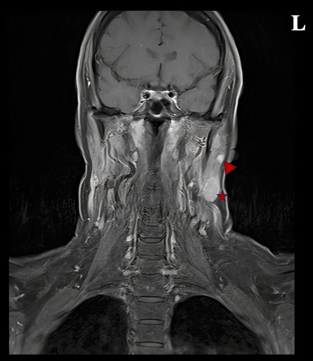
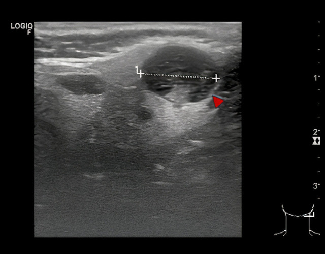
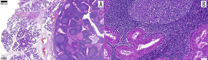
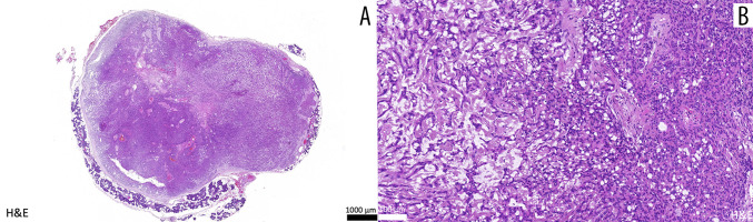
BACKGROUND Benign pleomorphic adenoma is the most common primary tumor of the salivary glands and mainly arises in the parotid gland. Warthin's tumor, or papillary cystadenoma lymphomatosum, represents <30% of benign parotid tumors. The simultaneous occurrence of multiple parotid tumors is rarely described - depending on the corresponding histology (different/identical), the time of their occurrence (synchronous/metachronous), as well as their location (unilateral/bilateral), multiple parotid tumors can be further sub-classified. CASE REPORT We describe the case of a 54-year-old female patient with progressive and painful swelling of the left parotid gland for the last 6 months. During extra-oral examination, a bulging, displaceable mass of approximately 3 cm was determined. A subsequent MRI (magnetic resonance imaging) examination revealed a multifocal lesion but failed to provide a decisive clue as to the tumor entity of the lesion, and a lateral (superficial) parotidectomy was performed. Postoperative histomorphological interpretation allowed the final pathological diagnosis of synchronous, unilateral occurrence of a pleomorphic adenoma as well as a Warthin's tumor. CONCLUSIONS This report presents a rare case of synchronous unilateral parotid tumors and supports that benign pleomorphic adenoma and Warthin's tumor are the most common associations. Since clinical examination, MRI imaging, and even cytological assessment could be misleading in the detection of synchronous ipsilateral multiple parotid gland tumors, our report also highlights the importance of timely and accurate diagnosis with histopathology to plan surgery and to exclude malignant transformation, which is a rare but important association with both types of primary salivary gland tumor.
Conflict of interest statement
Figures
Similar articles
-
Synchronous unilateral parotid neoplasms. A case report.Med Oral Patol Oral Cir Bucal. 2009 Feb 1;14(2):E90-2. Med Oral Patol Oral Cir Bucal. 2009. PMID: 19179956
-
[Synchronous double tumors of the parotid gland].Laryngorhinootologie. 1996 Jul;75(7):437-40. doi: 10.1055/s-2007-997610. Laryngorhinootologie. 1996. PMID: 8924174 German.
-
Multiple synchronous bilateral Warthin's tumors of the parotid glands with pleomorphic adenoma. Case report and review of the literature.Oral Surg Oral Med Oral Pathol. 1993 Sep;76(3):319-24. doi: 10.1016/0030-4220(93)90260-b. Oral Surg Oral Med Oral Pathol. 1993. PMID: 8397362 Review.
-
Synchronous oncocytoma and Warthin's tumor in the ipsilateral parotid gland.Auris Nasus Larynx. 2004 Mar;31(1):73-8. doi: 10.1016/j.anl.2003.07.008. Auris Nasus Larynx. 2004. PMID: 15041058
-
[Bilateral adenolymphoma (Warthin's tumor) of the parotid. The anatomicoclinical, diagnostic and therapeutic aspects of 2 cases].Minerva Stomatol. 1994 Nov;43(11):535-42. Minerva Stomatol. 1994. PMID: 7739487 Review. Italian.
Cited by
-
Eleven-Year Incidence of Salivary Gland Tumors-A Retrospective, Single-Centered Study in Croatia.Clin Pract. 2025 May 29;15(6):104. doi: 10.3390/clinpract15060104. Clin Pract. 2025. PMID: 40558222 Free PMC article.
References
-
- Speight P, Barrett A. Salivary gland tumours. Oral Dis. 2002;8(5):229–40. - PubMed
-
- Warthin AS. Papillary cyst adenoma lymphomatosum: A rare teratoid of the parotid region. J Cancer Res. 1929;13(2):116–25.
-
- WHO Classification of Tumours Editorial Board . Head and neck tumours. 5th edition. Lyon: International Agency for Research on Cancer; 2022.
-
- Bradley PJ, McGurk M. Incidence of salivary gland neoplasms in a defined UK population. Br J Oral Maxillofac Surg. 2013;51(5):399–403. - PubMed
-
- El-Naggar AK, Chan JKC, Grandis JR, et al., editors. WHO Classification of Head and Neck Tumours. Lyon: International Agency for Research on Cancer; 2017.
Publication types
MeSH terms
Supplementary concepts
LinkOut - more resources
Full Text Sources
