Epidermolysis bullosa in a mother-infant dyad
- PMID: 38033404
- PMCID: PMC10686001
- DOI: 10.1093/omcr/omad124
Epidermolysis bullosa in a mother-infant dyad
Abstract
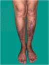
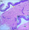
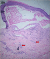
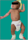
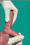
Epidermolysis Bullosa is an inherited mechanobullous disorder which presents in the neonatal period as blistering skin lesions. In this case report, we describe an uncommon presentation of Epidermolysis Bullosa Simplex in a term infant, weighing 2640 g, born to a mother who was also diagnosed with Epidermolysis Bullosa Pruriginosa during the course of the evaluation of her newborn. The clinical situation presented us with a unique dilemma with regard to routine newborn care practices including handling, skin and diaper care. Though the presentation was typically characteristic of EB, we illustrate with images the diagnostic modalities and challenges faced in the hospital while caring for this fragile skin in a low and middle-income country's neonatal intensive care unit. This is the first reported case of a neonate with Epidermolysis Bullosa Simplex born to a mother with Epidermolysis Bullosa Pruriginosa.
© The Author(s) 2023. Published by Oxford University Press.
Conflict of interest statement
None.
Figures



References
-
- Maldonado-Colin G, Hernández-Zepeda C, Durán-McKinster C, García-Romero MT. Inherited epidermolysis bullosa: a multisystem disease of skin and mucosae fragility. Indian J Paediat Dermatol 2017;18:267–73.
-
- Has C, Bauer JW, Bodemer C, Bolling MC, Bruckner-Tuderman L, Diem A. et al. Consensus reclassification of inherited epidermolysis bullosa and other disorders with skin fragility. Br J Dermatol 2020;183:614–27. - PubMed
-
- Lucky AW, Whalen J, Rowe S, Marathe KS, Gorell E. Diagnosis and Care of the Newborn with epidermolysis bullosa. NeoReviews 2021;22:e438–51. - PubMed
-
- Yiasemides E, Walton J, Marr P, Villanueva EV, Murrell DF. A comparative study between transmission electron microscopy and immunofluorescence mapping in the diagnosis of epidermolysis bullosa. Am J Dermatopathol 2006;28:387–94. - PubMed
-
- Sathishkumar D, Jacob AR. Learning to pop blisters in epidermolysis bullosa with a simple model. Pediatr Dermatol 2020;37:1215–7. - PubMed
Publication types
LinkOut - more resources
Full Text Sources

