. 2023 Nov 15:11:1294541.
doi: 10.3389/fcell.2023.1294541. eCollection 2023.

The relationship between miR-21, DNA methylation, and bisphenol a in bovine COCs and granulosa cells

Affiliations

The relationship between miR-21, DNA methylation, and bisphenol a in bovine COCs and granulosa cells

Reem Sabry et al. Front Cell Dev Biol. .

Abstract

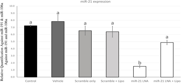
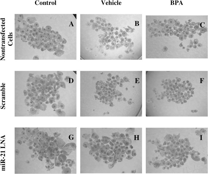
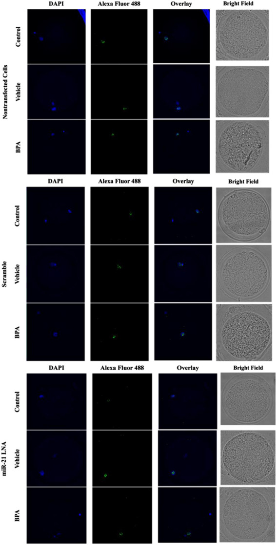
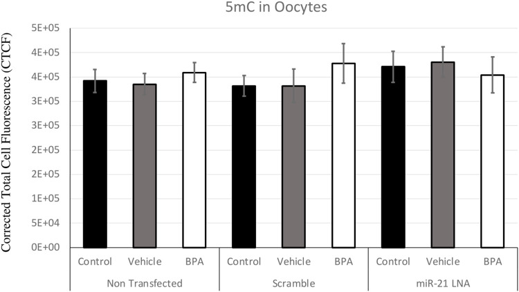
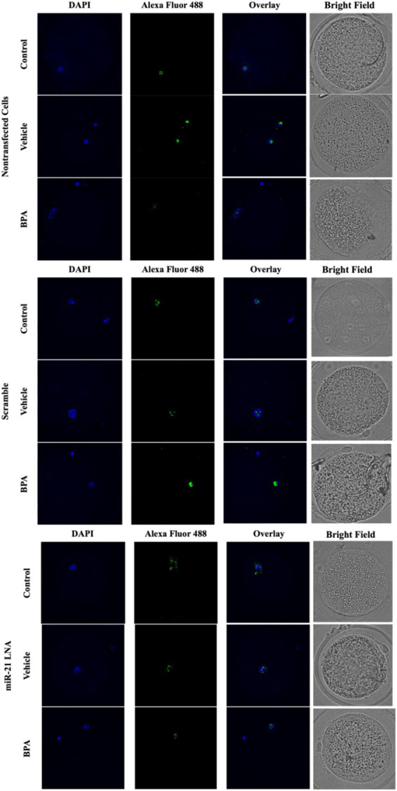
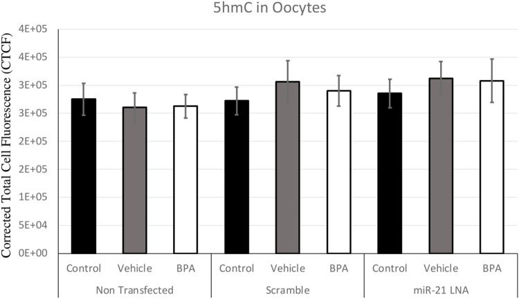
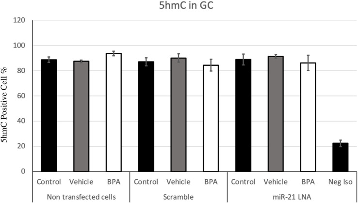
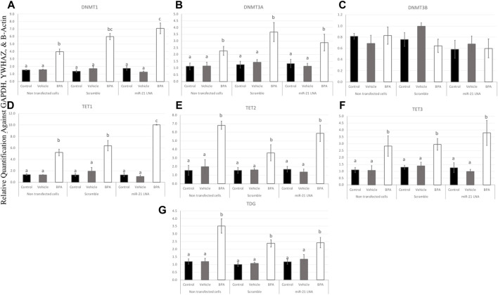
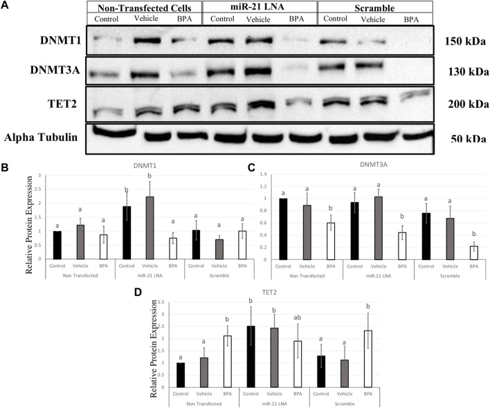
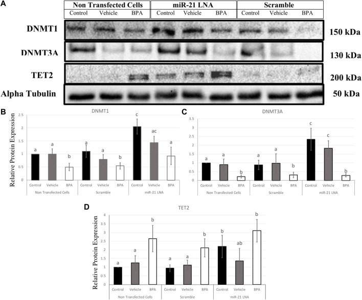
Introduction: miR-21 is a critical microRNA for the regulation of various processes in oocytes and granulosa cells. It is involved in the modulation of apoptosis and can influence other epigenetic mechanisms. Among these mechanisms, DNA methylation holds significant importance, particularly during female gametogenesis. Evidence has demonstrated that microRNAs, including miR-21, can regulate DNA methylation. Bisphenol A (BPA) is a widespread chemical that disrupts oocyte maturation and granulosa cell function. Recent findings suggested that BPA can act through epigenetic pathways, including DNA methylation and microRNAs. Methods: This study uses anti-miR-21 LNAs to explore the involvement of miR-21 in the regulation of DNA methylation in bovine Cumulus-Oocyte-Complexes (COCs) and granulosa cells, in the presence and absence of BPA. This study investigated 5 mC/5hmC levels as well as gene expression of various methylation enzymes using qPCR and western blotting. Results and discussion: Results reveal that BPA reduces 5mC levels in granulosa cells but not in COCs, which can be attributed to a decrease in the methylating enzymes DNMT1 and DNMT3A, and an increase in the demethylating enzyme TET2. We observed a significant increase in the protein levels of DNMT1, DNMT3A, and TET2 upon inhibition of miR-21 in both COCs and granulosa cells. These findings directly imply a strong correlation between miR-21 signaling and the regulation of DNA methylation in bovine COCs and granulosa cells under BPA exposure.

Keywords: DNA methylation; bisphenol A; granulosa cells; miR-21; oocytes.


Conflict of interest statement

The authors declare that the research was conducted in the absence of any commercial or financial relationships that could be construed as a potential conflict of interest.

Figures

FIGURE 1
FIGURE 1
miR-21 expression in in vitro matured bovine COCs. Bovine COCs were matured in in vitro S-IVM media and treated with either an LNA scramble or LNA inhibitor in the presence and absence of Lipofectamine 3,000 at 0.1 µM and 0.5 µM, respectively. Results show that treatment with the miR-21 inhibitor alone resulted in an 80% knockdown of miR-21 expression. Different letters indicate significant differences, with b indicating a significantly different mean than a at p < 0.05. Bars represent the mean ± SEM.
FIGURE 2
FIGURE 2
Oocyte Morphology after 24 h in vitro maturation with anti-miR-21 LNAs and BPA. Bovine COCs were placed into maturation media alone (A–C), in the presence of a scramble (D–F), or in the presence of anti-miR-21 LNA inhibitors (G–I) at 0.5 µM. 12 h into maturation, COCs were also treated with either a vehicle (B,E,H) or BPA (C,F,I) at 0.05 mg/mL for another 12 h. BPA-treated COCs appear poorer in quality with less cumulus cell expansion and increased heterogeneity among COC phenotypes. Scale bar = 500 µm.
FIGURE 3
FIGURE 3
Confocal Microscopy of 5′methylcytosine in Bovine Oocytes. Transfected and treated COCs were fixed and permeabilized to allow for immunostaining of 5 mC. Immunodetection of 5 mC in COCs was achieved by utilizing an Alexa Fluor 488 conjugated secondary antibody coupled with confocal microscopy. Confocal Images are represented for all 9 groups.
FIGURE 4
FIGURE 4
Corrected Total Cell Fluorescence of 5′methylcytosine in Bovine Oocytes. Transfected and treated COCs were fixed and permeabilized to allow for immunostaining of 5 mC. The corrected total cell fluorescence was calculated using ImageJ on a minimum of 15 COCs per group for a total of 140 COCs analyzed. Bars represent the mean ± SEM at p < 0.05.
FIGURE 5
FIGURE 5
Confocal Microscopy of 5′hydroxymethylcytosine in Bovine Oocytes. Transfected and treated COCs were fixed and permeabilized to allow for immunostaining of 5 hmC. Immunodetection of 5 hmC in COCs was also achieved by utilizing an Alexa Fluor 488 conjugated secondary antibody coupled with confocal microscopy. Confocal Images are represented for all 9 groups.
FIGURE 6
FIGURE 6
Corrected Total Cell Fluorescence of 5′hydroxymethylcytosine in Bovine Oocytes. Transfected and treated COCs were fixed and permeabilized to allow for immunostaining of 5 hmC. The corrected total cell fluorescence was calculated using ImageJ on a minimum of 15 COCs per group for a total of 140 COCs analyzed. Bars represent the mean ± SEM at p < 0.05.
FIGURE 7
FIGURE 7
Quantification of 5′methylcytosine (5 mC) in Bovine Granulosa Cells. Analysis of 5 mC levels was done on FlowJo on a minimum of three biological replicates. The last column represents the negative control where a negative isotype was used as the primary antibody. Different letters indicate significant differences, with b indicating a significantly different mean than a at p < 0.05. Bars represent the mean ± SEM and n = 7.
FIGURE 8
FIGURE 8
Quantification of 5′hydroxymethylcytosine (5 hmC) in Bovine Granulosa Cells. The analysis was done on FlowJo on a minimum of three biological replicates. The last column represents the negative control where a negative isotype was used as the primary antibody. Bars represent the mean ± SEM at p < 0.05and n = 4.
FIGURE 9
FIGURE 9
Expression of methylating and demethylating mRNAs after miR-21 inhibition and BPA treatment in Bovine COCs. COCs were transfected with LNA inhibitor probes at 0.5 μM then treated with BPA (0.05 mg/mL). All transcripts including DNMT1 (A), DNMT3A (B), TET1—3 (D–F), and TDG (G) were significantly increased after BPA exposure in all transfection groups apart from DNMT3B (C). Quantification is normalized to reference targets GAPDH, YWHAZ, and B-Actin. Different letters indicate significant differences, with b indicating a significantly different mean than a at p < 0.05, c indicating a significantly different mean than a and b, and d indicating a significantly different mean than a, b, and c at p < 0.05. ab indicates no differences between a or b and bc indicates no differences between b or c. Bars represent the mean ± SEM.
FIGURE 10
FIGURE 10
Expression of methylating and demethylating mRNAs after miR-21 inhibition and BPA treatment in Bovine GCs. Cells were transfected with LNA inhibitor probes at 0.5 μM for 12 h and then treated with BPA (0.05 mg/mL) for another 12 h. Most transcripts including DNMT1 (A), DNMT3A (B), TET3 (F), and TDG (G) were significantly increased after BPA exposure in all transfection groups apart from TET1 (D) that was decreased, DNMT3B (C) that was unaffected, and TET2 (E) that was unaffected in the inhibited group. Quantification is normalized to reference targets GAPDH, YWHAZ, and B-Actin. Different letters indicate significant differences, with b indicating a significantly different mean than a at p < 0.05. Bars represent the mean ± SEM.
FIGURE 11
FIGURE 11
Relative protein expression of DNMT1, DNMT3A, and TET2 after miR-21 inhibition and BPA treatment in Bovine COCs. Transfections were done with LNA inhibitor probes at 0.5 μM for 12 h followed by BPA treatment for another 12 h. Western blots (A) and graphical representations of DNMT1 (B), DNMT3A (C), and TET2 (D) revealed that BPA decreased and increased DNMT3A and TET2 proteins, respectively. miR-21 Inhibition also induced protein expression in the control-only groups for DNMT1 and TET2. Densitometric analysis was performed relative to the loading control, α-tubulin. Different letters indicate significant differences, with b indicating a significantly different mean than a at p < 0.05. Bars represent the mean ± SEM. n = 6 for DNMT1; n = 9 for DNMT3A; n = 6 for TET2.
FIGURE 12
FIGURE 12
Relative protein expression of DNMT1, DNMT3A, and TET2 after miR-21 inhibition and BPA treatment in Bovine GCs. Transfections were done with LNA inhibitor probes at 0.5 μM for 12 h followed by BPA treatment for another 12 h. Western blots (A) and graphical representations of DNMT1 (B), DNMT3A (C), and TET2 (D) revealed that BPA decreased and increased DNMTs and TET2 proteins, respectively. miR-21 Inhibition also induced all protein expression in the control-only groups. Densitometric analysis was performed relative to the loading control, α-tubulin. Different letters indicate significant differences, with b indicating a significantly different mean than a at p < 0.05. Bars represent the mean ± SEM. n = 9 for DNMT1, n = 6 for DNMT3A, n = 6 for TET2.

Similar articles

Cited by

References

    1. Aldakheel F. M., Abuderman A. A., Alduraywish S. A., Xiao Y., Guo W. W. (2021). MicroRNA-21 inhibits ovarian granulosa cell proliferation by targeting SNHG7 in premature ovarian failure with polycystic ovary syndrome. J. Reprod. Immunol. 146, 103328. 10.1016/J.JRI.2021.103328 - DOI - PubMed
    1. Awada Z., Nasr R., Akika R., Cahais V., Cuenin C., Zhivagui M., et al. (2019). DNA methylome-wide alterations associated with estrogen receptor-dependent effects of bisphenols in breast cancer. Clin. Epigenetics 11, 138–217. 10.1186/s13148-019-0725-y - DOI - PMC - PubMed
    1. Bartolucci A. F., Uliasz T., Peluso J. J. (2020). MicroRNA-21 as a regulator of human cumulus cell viability and its potential influence on the developmental potential of the oocyte. Biol. Reprod. 103, 94–103. 10.1093/BIOLRE/IOAA058 - DOI - PubMed
    1. Bhandari R. K., Taylor J. A., Sommerfeld-Sager J., Tillitt D. E., Ricke W. A., Vom Saal F. S. (2019). Estrogen receptor 1 expression and methylation of Esr1 promoter in mouse fetal prostate mesenchymal cells induced by gestational exposure to bisphenol A or ethinylestradiol. Environ. Epigenet 5, dvz012–8. 10.1093/EEP/DVZ012 - DOI - PMC - PubMed
    1. Bhaskaran M., Mohan M. (2014). MicroRNAs: history, biogenesis, and their evolving role in animal development and disease. Vet. Pathol. 51, 759–774. 10.1177/0300985813502820 - DOI - PMC - PubMed

LinkOut - more resources