Case Reports
. 2023 Oct 23:13:e2023448.
doi: 10.4322/acr.2023.448. eCollection 2023.

Radiologic-pathologic autopsy correlation of an internal watershed infarct, a case report

Affiliations
Case Reports

Radiologic-pathologic autopsy correlation of an internal watershed infarct, a case report

Thomas Dimitrios Zaikos et al. Autops Case Rep. .

Abstract

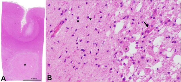
Internal watershed infarcts (IWIs) occur at the junction of the deep and superficial perforating arterial branches of the cerebrum. Despite documentation in the radiology literature, IWIs are rarely encountered at the time of autopsy. Here, we report the case of a 59-year-old incarcerated male who was brought to the emergency department after being found unresponsive on the floor of his jail cell. Initial examination and imaging demonstrated right-sided hemiplegia, aphasia, right facial droop, and severe stenosis of the left middle cerebral artery, respectively. Repeat imaging 4 days after admission and 26 days before death demonstrated advanced stenosis of the intracranial, communicating segment of the right internal carotid artery, a large acute infarct in the right posterior cerebral artery territory, and bilateral deep white matter ischemic changes with a right-sided "rosary-like" pattern of injury that is typical of IWIs. Postmortem gross examination showed that the right deep white matter lesion had progressed to a confluent, "cigar-shaped" subacute IWI involving the right corona radiata. This is the first well-documented case of an IWI with radiologic imaging and photographic gross pathology correlation. This case uniquely highlights a rarely encountered lesion at the time of autopsy and provides an excellent visual representation of internal watershed neuroanatomy.

Keywords: Internal watershed; autopsy; infarction; neuropathology.


Conflict of interest statement

Conflict of interest: None.

Figures

Figure 1
Figure 1. Magnetic resonance imaging from hospital day 4. A - the acute large right posterior cerebral artery infraction is seen affecting the right medial temporal lobe and occipital lobe on the diffusion weighted imaging; B - at the upper border of the right parieto-occipital cortical infarction, one can see a string of bright areas in the deep centrum semiovale on the right side representing acute watershed infarction on this diffusion weighted image. Acute cortical strokes are seen in the left hemisphere; C - the fluid attenuated inversion recovery (FLAIR) scan confirms the deep watershed infarctions bilaterally, seen as higher signal intensity on this imaging. The visualization on FLAIR suggests the infarctions are over 6 hours old. Diffusion weighted imaging remain positive for 7-15 days.
Figure 2
Figure 2. A and B - gross and microscopic pathology of internal watershed infarct; A - a confluent, “cigar-shaped” lesion involving the right hemispheric deep white matter was identified at the time of brain cutting (red arrow). This lesion extended from the frontal pole to the parieto-occipital lobe.
Figure 3
Figure 3. Microphotographs of the Brain - A - low-magnification photograph of representative section of the lesion (asterisk) and overlying cortex (section taken from area of red box from panel A); B - representative histology of the interface between the lesion demonstrating a necrotic core with infiltrating macrophages (asterisk) and neovascularization (arrow) as well as adjacent reactive glial tissue (arrowheads) (A - H&E; B - H&E, 200x).

References

    1. Bladin CF, Chambers BR. Clinical features, pathogenesis, and computed tomographic characteristics of internal watershed infarction. Stroke. 1993;24(12):1925–1932. doi: 10.1161/01.STR.24.12.1925. - DOI - PubMed
    1. Bogousslavsky J. The plurality of subcortical infarction. Stroke. 1992;23(5):629–631. doi: 10.1161/01.STR.23.5.629. - DOI - PubMed
    1. Van Den Bergh R. Centrifugal elements in the vascular pattern of the deep intracerebral blood supply. Angiology. 1969;20(2):88–94. doi: 10.1177/000331976902000205. - DOI - PubMed
    1. Moody DM, Bell MA, Challa VR. Features of the cerebral vascular pattern that predict vulnerability to perfusion or oxygenation deficiency: an anatomic study. AJNR Am J Neuroradiol. 1990;11(3):431–439. - PMC - PubMed
    1. Momjian-Mayor I, Baron JC. The pathophysiology of watershed infarction in internal carotid artery disease: review of cerebral perfusion studies. Stroke. 2005;36(3):567–577. doi: 10.1161/01.STR.0000155727.82242.e1. - DOI - PubMed

Publication types