

Expression of the kidney anion exchanger 1 affects WNK4 and SPAK phosphorylation and results in claudin-4 phosphorylation
- PMID: 38034706
- PMCID: PMC10687047
- DOI: 10.1016/j.heliyon.2023.e22280
Expression of the kidney anion exchanger 1 affects WNK4 and SPAK phosphorylation and results in claudin-4 phosphorylation
Abstract
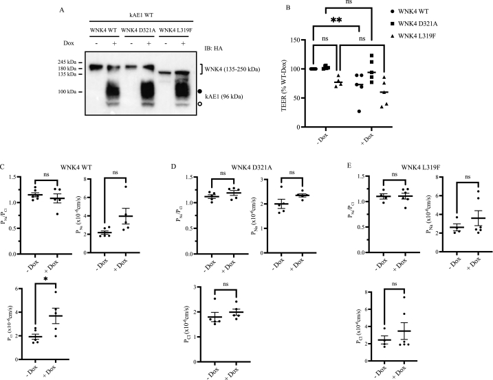
In the renal collecting ducts, chloride reabsorption occurs through both transcellular and paracellular pathways. Recent literature highlights a functional interplay between both pathways. We recently showed that in polarized inner medullary collecting duct cells, expression of the basolateral kidney anion exchanger 1 (kAE1) results in a decreased transepithelial electrical resistance (TEER), in a claudin-4 dependent pathway. Claudin-4 is a paracellular sodium blocker and chloride pore. Here, we show that kAE1 expression in mouse inner medullary collecting duct cells triggers WNK4, SPAK and claudin-4 phosphorylation. Expression of a functionally dead kAE1 E681Q mutant has no effect on phosphorylation of these proteins. Expression of a catalytically inactive WNK4 D321A or chloride-insensitive WNK4 L319F mutant abolishes kAE1 effect on TEER, supporting a contribution of WNK4 to the process. We propose that variations of the cytosolic pH and chloride concentration upon kAE1 expression alter WNK4 kinase activity and tight junction properties.
Keywords: Blood pressure; Chloride conservation; Claudin; Collecting duct; Distal nephron; Epithelium; Intercalated cells; Kidney; Membrane protein; Paracellular proteins; Sodium conservation; Tight junctions; Transporters.
© 2023 Published by Elsevier Ltd.
Conflict of interest statement
The authors declare the following financial interests/personal relationships which may be considered as potential competing interests: Emmanuelle Cordat reports financial support was provided by 10.13039/501100000024Canadian Institutes of Health Research. Rawad Lashhab reports financial support was provided by 10.13039/501100000038Natural Sciences and Engineering Research Council of Canada. Maria Chavez-Canales reports financial support was provided by 10.13039/501100003141CONACyT Mexicon. Maria Chavez-Canales reports financial support was provided by PAPIIT UNAM.
Figures


Similar articles
-
The kidney anion exchanger 1 affects tight junction properties via claudin-4.Sci Rep. 2019 Feb 28;9(1):3099. doi: 10.1038/s41598-019-39430-9. Sci Rep. 2019. PMID: 30816203 Free PMC article.
-
Claudin-4 forms paracellular chloride channel in the kidney and requires claudin-8 for tight junction localization.Proc Natl Acad Sci U S A. 2010 Oct 19;107(42):18010-5. doi: 10.1073/pnas.1009399107. Epub 2010 Oct 4. Proc Natl Acad Sci U S A. 2010. PMID: 20921420 Free PMC article.
-
Interaction between Epithelial Sodium Channel γ-Subunit and Claudin-8 Modulates Paracellular Sodium Permeability in Renal Collecting Duct.J Am Soc Nephrol. 2020 May;31(5):1009-1023. doi: 10.1681/ASN.2019080790. Epub 2020 Apr 3. J Am Soc Nephrol. 2020. PMID: 32245797 Free PMC article.
-
Regulation of paracellular transport in the distal nephron.Curr Opin Nephrol Hypertens. 2012 Sep;21(5):547-51. doi: 10.1097/MNH.0b013e328355cb47. Curr Opin Nephrol Hypertens. 2012. PMID: 22691877 Free PMC article. Review.
-
Paracellular transport in the collecting duct.Curr Opin Nephrol Hypertens. 2016 Sep;25(5):424-8. doi: 10.1097/MNH.0000000000000253. Curr Opin Nephrol Hypertens. 2016. PMID: 27490784 Free PMC article. Review.
Cited by
-
Urinary sodium wasting and disrupted collecting duct function in mice with distal renal tubular acidosis mutations.Dis Model Mech. 2025 May 1;18(5):dmm052138. doi: 10.1242/dmm.052138. Epub 2025 May 23. Dis Model Mech. 2025. PMID: 40289527 Free PMC article.
References
-
- Kiuchi-Saishin Y., Gotoh S., Furuse M., Takasuga A., Tano Y., Tsukita S. Differential expression patterns of claudins, tight junction membrane proteins, in mouse nephron segments. J. Am. Soc. Nephrol. 2002;13:875–886. http://www.ncbi.nlm.nih.gov/pubmed/11912246 - PubMed
LinkOut - more resources
Full Text Sources
