

Spatial distributions of white matter hyperintensities on brain MRI: A pooled analysis of individual participant data from 11 memory clinic cohorts
- PMID: 38035457
- PMCID: PMC10698002
- DOI: 10.1016/j.nicl.2023.103547
Spatial distributions of white matter hyperintensities on brain MRI: A pooled analysis of individual participant data from 11 memory clinic cohorts
Abstract
Introduction: The spatial distribution of white matter hyperintensities (WMH) on MRI is often considered in the diagnostic evaluation of patients with cognitive problems. In some patients, clinicians may classify WMH patterns as "unusual", but this is largely based on expert opinion, because detailed quantitative information about WMH distribution frequencies in a memory clinic setting is lacking. Here we report voxel wise 3D WMH distribution frequencies in a large multicenter dataset and also aimed to identify individuals with unusual WMH patterns.
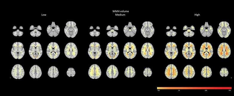
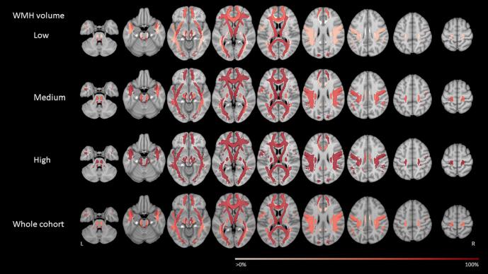
Methods: Individual participant data (N = 3525, including 777 participants with subjective cognitive decline, 1389 participants with mild cognitive impairment and 1359 patients with dementia) from eleven memory clinic cohorts, recruited through the Meta VCI Map Consortium, were used. WMH segmentations were provided by participating centers or performed in Utrecht and registered to the Montreal Neurological Institute (MNI)-152 brain template for spatial normalization. To determine WMH distribution frequencies, we calculated WMH probability maps at voxel level. To identify individuals with unusual WMH patterns, region-of-interest (ROI) based WMH probability maps, rule-based scores, and a machine learning method (Local Outlier Factor (LOF)), were implemented.
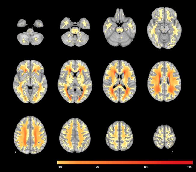
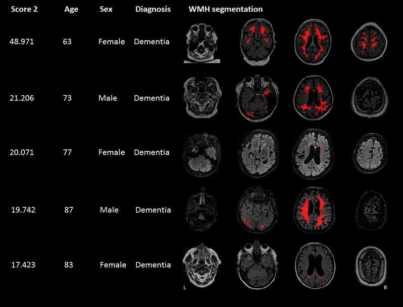
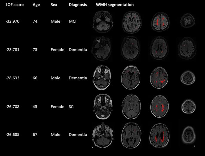
Results: WMH occurred in 82% of voxels from the white matter template with large variation between subjects. Only a small proportion of the white matter (1.7%), mainly in the periventricular areas, was affected by WMH in at least 20% of participants. A large portion of the total white matter was affected infrequently. Nevertheless, 93.8% of individual participants had lesions in voxels that were affected in less than 2% of the population, mainly located in subcortical areas. Only the machine learning method effectively identified individuals with unusual patterns, in particular subjects with asymmetric WMH distribution or with WMH at relatively rarely affected locations despite common locations not being affected.
Discussion: Aggregating data from several memory clinic cohorts, we provide a detailed 3D map of WMH lesion distribution frequencies, that informs on common as well as rare localizations. The use of data-driven analysis with LOF can be used to identify unusual patterns, which might serve as an alert that rare causes of WMH should be considered.
Keywords: Brain MRI; Distribution frequencies; Lesion location; White matter hyperintensities.
Copyright © 2023 The Authors. Published by Elsevier Inc. All rights reserved.
Conflict of interest statement
Declaration of competing interest The authors declare the following financial interests/personal relationships which may be considered as potential competing interests: FB is supported by the NIHR biomedical research center at UCLH. MD received honoraria for lectures from Bayer Vital and Sanofi Genzyme, Consultant for Hovid Berhad and Roche Pharma. RWP received honoraria from GE Healthcare and is co-lead of Neurofilament light consortium. CHS is supported by an Alzheimer's Society Fellowship. The remaining authors have nothing to disclose.
Figures
References
-
- Auer DP, Pütz B, Gössl C, Elbel GK, Gasser T, Dichgans M. Differential Lesion Patterns in CADASIL and Sporadic Subcortical Arteriosclerotic Encephalopathy: MR Imaging Study with Statistical Parametric Group Comparison. Vol 218.; 2001. - PubMed
-
- Barkhof F., Scheltens P. Imaging of white matter lesions. Cerebrovasc Dis. 2002;13(suppl 2):21–30. - PubMed
