. 2023 Nov 30;14(1):7908.
doi: 10.1038/s41467-023-43657-6.

Discovery of a Drug-like, Natural Product-Inspired DCAF11 Ligand Chemotype

Affiliations

Discovery of a Drug-like, Natural Product-Inspired DCAF11 Ligand Chemotype

Gang Xue et al. Nat Commun. .

Abstract

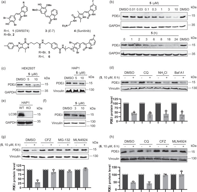
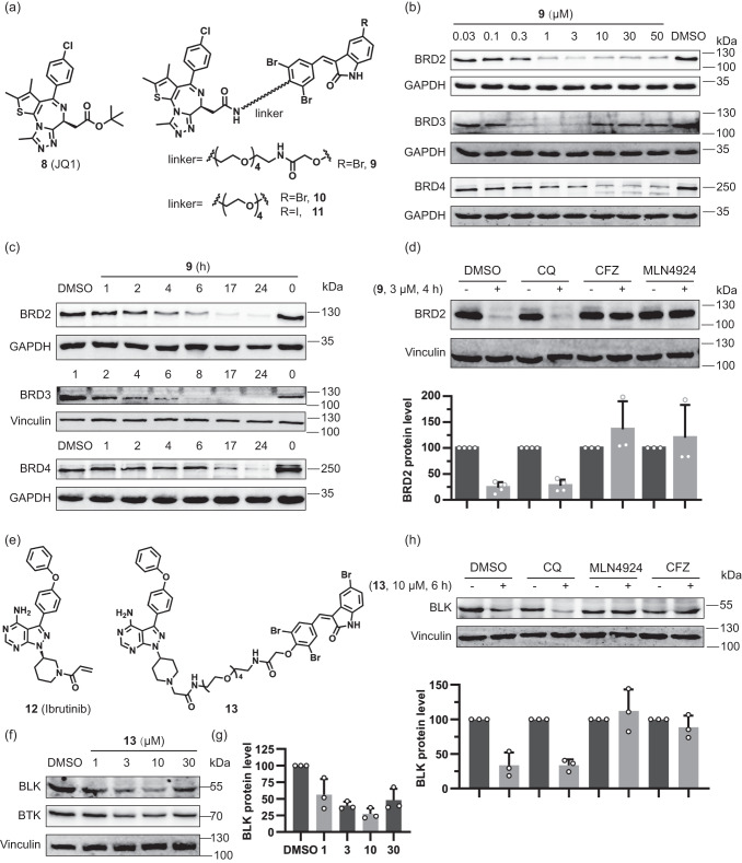
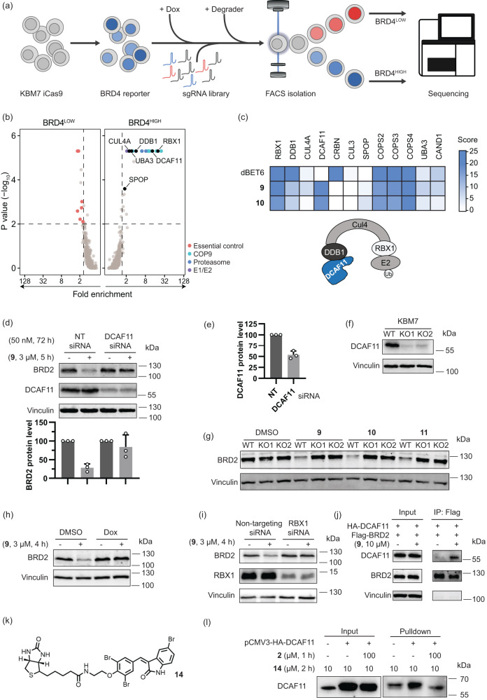
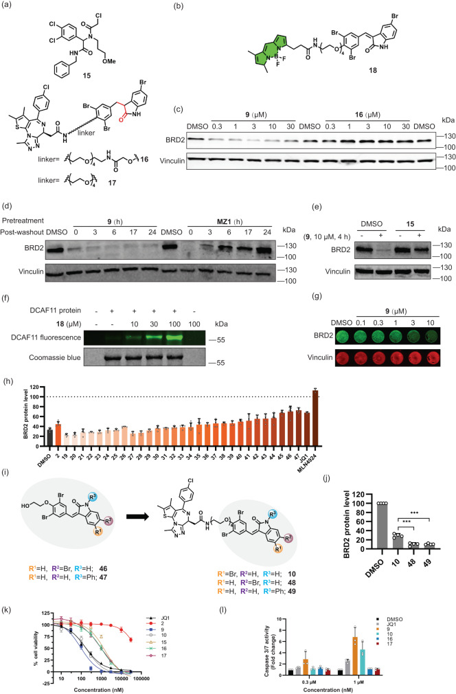
Targeted proteasomal and autophagic protein degradation, often employing bifunctional modalities, is a new paradigm for modulation of protein function. In an attempt to explore protein degradation by means of autophagy we combine arylidene-indolinones reported to bind the autophagy-related LC3B-protein and ligands of the PDEδ lipoprotein chaperone, the BRD2/3/4-bromodomain containing proteins and the BTK- and BLK kinases. Unexpectedly, the resulting bifunctional degraders do not induce protein degradation by means of macroautophagy, but instead direct their targets to the ubiquitin-proteasome system. Target and mechanism identification reveal that the arylidene-indolinones covalently bind DCAF11, a substrate receptor in the CUL4A/B-RBX1-DDB1-DCAF11 E3 ligase. The tempered α, β-unsaturated indolinone electrophiles define a drug-like DCAF11-ligand class that enables exploration of this E3 ligase in chemical biology and medicinal chemistry programs. The arylidene-indolinone scaffold frequently occurs in natural products which raises the question whether E3 ligand classes can be found more widely among natural products and related compounds.


Conflict of interest statement

G.E.W. is scientific founder and shareholder of Proxygen and Solgate. The Winter lab received research funding from Pfizer. The remaining co-authors declare no conflicts of interest to disclose.

Figures

Fig. 1
Fig. 1. Degradation of PDEδ induced by bifunctional arylidene-indolinones 5 and 6.
a Structures of selected indolinones and bifunctional degraders derived therefrom. b Dose-dependent degradation of PDEδ in Jurkat cells treated with compound 5 (6 h) and time-dependent degradation of PDEδ in Jurkat cells treated with compound 5 (10 μM). Representative result of n = 3. c PDEδ levels in HEK293T and HAP1 cells treated with compound 5 at 5 μM or 10 μM. Representative result of n = 3. d PDEδ levels in Jurkat cells pretreated with autophagy inhibitors Chloroquine (CQ, 50 μM), NH4Cl (15 mM), and Bafilomycin A1 (Baf A1, 0.3 μM) for 2 h prior to the addition of 10 µM compound 5 for another 6 h. Quantification of the relative PDEδ protein content in relation to the DMSO control is shown in the bar graph. Data are mean values ± SD (n = 5 biological replicates). e, f PDEδ levels in LC3B knockout HAP1 cells. Representative result of n = 3. e Lack of LC3B protein in LC3B knockout HAP1 cells. f LC3B knockout HAP1 cells were treated with compound 5, and PDEδ levels were analyzed using immunoblotting. g PDEδ levels in Jurkat cells pretreated with the proteasome inhibitors Carfilzomib (CFZ, 0.2 μM) or MG-132 (10 μM), or the neddylation inhibitor MLN4924 (1 μM) for 2 h, followed by treatment with 10 µM of compound 5 for another 6 h. Quantification of the relative PDEδ protein content in relation to the DMSO control is shown in the bar graph. Data are mean values ± SD (n = 3 biological replicates). h Western blots of PDEδ in Jurkat cells pretreated with CQ (50 μM), CFZ (0. 2 μM) or MLN4924 (1 μM) for 2 h, followed by treatment with 10 µM compound 6 for another 6 h. Quantification of the relative PDEδ protein content in relation to the DMSO control is shown in the bar graph. Data are mean values ± SD (n = 3 biological replicates).
Fig. 2
Fig. 2. Degradation activity and mechanism of action of bifunctional compounds that target BET proteins or BTK/BLK.
a Structures of the BET inhibitor JQ1 and selected bifunctional degraders. b Dose-dependent degradation of BET proteins in Jurkat cells treated with compound 9 for 6 h. Representative result of n = 3 is shown. c Time-dependent degradation of BET proteins in Jurkat cells treated with compound 9 at 3 μM. Representative result of n = 3. d BRD2 protein levels in Jurkat cells pretreated with the autophagy inhibitor CQ (50 μM), proteasome inhibitor CFZ (1 μM) and neddylation inhibitor MLN4924 (1 μM) for 40 min, followed by treatment with 3 µM compound 9 for another 4 h. Quantification of the relative BRD2 protein content in relation to the DMSO control is shown in the bar graph. Data are mean values ± SD (n = 4 biological replicates for DMSO and CQ; n = 3 biological replicates for MLN4924 and CFZ). e Structures of the BTK- and BLK inhibitor ibrutinib and related bifunctional degrader. f Dose-dependent degradation of BTK- and BLK proteins in Jurkat cells treated with compound 13 for 6 h. Representative result of n = 3 is shown. g Quantification of the relative BLK protein content, which was normalized to the DMSO control. Data are mean values ± SD (n = 3 biological replicates). h BLK protein levels in Ramos cells pretreated with the autophagy inhibitor CQ (50 μM), proteasome inhibitor CFZ (1 μM) and neddylation inhibitor MLN4924 (1 μM) for 40 min, followed by treatment with 10 µM of compound 13 for another 6 h. Quantification of the relative BLK protein content in relation to the DMSO control is shown in the bar graph. Data are mean values ± SD (n = 3 biological replicates).
Fig. 3
Fig. 3. Compound 9 promotes proteasomal degradation of BET proteins via the CRLDCAF11 complex.
a Schematic of FACS-based BRD4 protein stability CRISPR screen assay. b, c Identification of genes required for compound 9-mediated degradation of BRD4. b FACS-based CRISPR screens for regulators of BRD4 degradation induced by 9. Average gene-level fold-changes and non-adjusted one-sided p-values of BRD4HIGH and BRD4LOW cell populations compared to BRD4MID fraction were calculated using MAGeCK. Essential control genes (BRD4LOW) and 20S proteasome subunits, COP9 signalosome subunits and E1 or E2 ubiquitin ligases (BRD4HIGH) inside the scoring window (p-value < 0.01, fold-change > 1.5) are labelled. c Heatmap of selected screen hits from Fig. 3b and Supplementary Fig. 4a, b. Gene-level score was calculated as Log2 (fold change) * Log10 (p-value). d, e BRD2 and DCAF11 protein levels in Hep G2 cells transfected with non-targeting (NT) siRNA or DCAF11 siRNA, followed by 9 addition. d Quantification of the relative BRD2 levels. Data are mean values ± SD (n = 3 biological replicates). e Quantification of the relative DCAF11 levels. Data are mean values ± SD (n = 3 biological replicates). f DCAF11 levels in wild type (WT) KBM7 cells and DCAF11 knockout (KO1 and KO2) KBM7 cells. Representative result of n = 3. g BRD2 levels in WT and DCAF11 knockout (KO1 and KO2) KBM7 cells treated with compound 9 at 3 μM, 10 at 10 μM and 11 at 10 μM, for 6 h. Data are representative of n = 3. h BRD2 levels in Dox-inducible DDB1 knockout KBM7 cells pretreated with 0.5 µg/mL Dox for 3 days followed by addition of 9 at 3 μM for another 4 h. Representative result of n = 3. i BRD2 and RBX1 levels in MDA-MB-231 cells with NT or RBX1 siRNA followed by 9 addition. Representative result of n = 3. j BRD2 co-immunoprecipitation. FLAG-BRD2 protein was enriched by anti-FLAG beads and samples were analyzed for DCAF11. Representative result of n = 2. k Structure of biotinylated affinity probe 14. l Affinity-based enrichment by pulldown in Hep G2 cells overexpressing DCAF11. Protein levels were analyzed by WB. Representative result of n = 2.
Fig. 4
Fig. 4. Bifunctional compounds bind covalently to DCAF11 and display antiproliferative activity.
a Structures of 15 and bifunctional compounds 16 and 17 with saturated double bond. b Structure of probe 18 with BODIPY as fluorophore. c BRD2 levels in KBM7 cells treated with compounds 9 and 16 for 4 h. Representative result of n = 3. d BRD2 levels in Jurkat cells treated with PROTAC MZ1 or 9 (10 μM, 5 h) prior to washout and further incubation for the indicated time. Representative results of n = 3. e BRD2 levels in Hep G2 cells pretreated with 30 μM 15 for 1 h followed by 10 μM of compound 9 for another 4 h. Representative result of n = 2. f Fluorescence labelling of DCAF11. Purified DCAF11 protein was incubated with 18 for 40 min followed by analysis of in-gel fluorescence. Representative result of n = 3. g In-Cell Western for BRD2 levels in Hep G2 cells treated with 9 for 4 h. Representative results (n = 4 biological replicates). h Relative BRD2 protein levels as detected using In-Cell Western in Hep G2 cells pretreated with compounds 219-47 or JQ1 (structure shown in Supplementary Table 1) at 30 μM or MLN4924 at 1 μM for 1 h, followed by 9 (10 μM, 4 h) or DMSO addition. Data are mean values ± SD (n = 3 for JQ1 and MLN4924, n = 4 for 19–45, n = 5 for DMSO, 2, 46–47; n = biological replicates). i Structures of representative indolinones (46, 47) and corresponding bifunctional compounds (48, 49). j BRD2 levels in Jurkat cells treated with 9, 48 or 49 at 3 μM for 6 h. Data are mean values ± SD (n = 4 biological replicates). Statistical significance was calculated with unpaired two-tailed Student’s t-tests comparing 10 to 48 or 49 (P = 0.0002 or 0.0003 respectively; ***P < 0.001). k Influence on cell viability. Data are mean values ± SD (n = 3 biological replicates). The data of GI50 are shown in Supplementary Fig. 7h. l Caspase-3/7 activity for apoptosis detection. Data are mean values ± SD (n = 3 biological replicates).

References

    1. Domostegui A, Nieto-Barrado L, Perez-Lopez C, Mayor-Ruiz C. Chasing molecular glue degraders: screening approaches. Chem. Soc. Rev. 2022;51:5498–5517. doi: 10.1039/D2CS00197G. - DOI - PubMed
    1. Gerry CJ, Schreiber SL. Unifying principles of bifunctional, proximity-inducing small molecules. Nat. Chem. Biol. 2020;16:369–378. doi: 10.1038/s41589-020-0469-1. - DOI - PMC - PubMed
    1. Hanzl A, Winter GE. Targeted protein degradation: current and future challenges. Curr. Opin. Chem. Biol. 2020;56:35–41. doi: 10.1016/j.cbpa.2019.11.012. - DOI - PMC - PubMed
    1. Bekes M, Langley DR, Crews CM. PROTAC targeted protein degraders: the past is prologue. Nat. Rev. Drug Discov. 2022;21:181–200. doi: 10.1038/s41573-021-00371-6. - DOI - PMC - PubMed
    1. Cao C, He M, Wang L, He Y, Rao Y. Chemistries of bifunctional PROTAC degraders. Chem. Soc. Rev. 2022;51:7066–7114. doi: 10.1039/D2CS00220E. - DOI - PubMed

Publication types