. 2023 Nov 30;13(1):21142.
doi: 10.1038/s41598-023-48146-w.

Low-dose brain radiation: lowering hyperphosphorylated-tau without increasing DNA damage or oncogenic activation

Affiliations

Low-dose brain radiation: lowering hyperphosphorylated-tau without increasing DNA damage or oncogenic activation

Diego Iacono et al. Sci Rep. .

Abstract

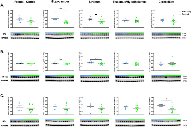
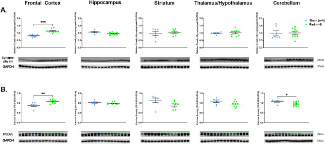
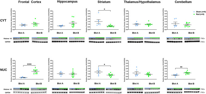
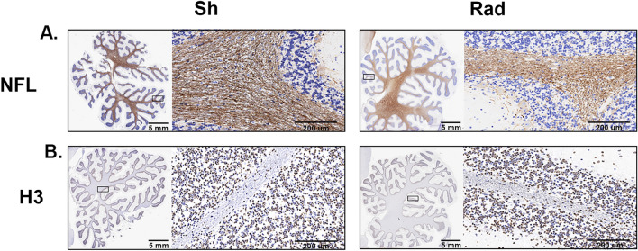
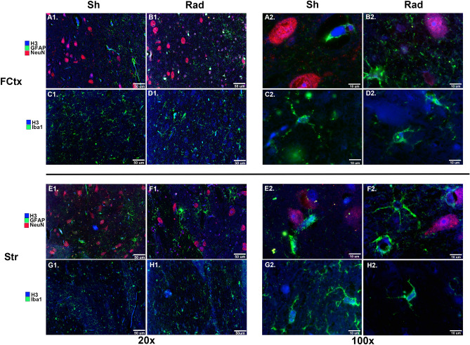
Brain radiation has been medically used to alter the metabolism of cancerous cells and induce their elimination. Rarely, though, brain radiation has been used to interfere with the pathomechanisms of non-cancerous brain disorders, especially neurodegenerative disorders. Data from low-dose radiation (LDR) on swine brains demonstrated reduced levels of phosphorylated-tau (CP13) and amyloid precursor protein (APP) in radiated (RAD) versus sham (SH) animals. Phosphorylated-tau and APP are involved in Alzheimer's disease (AD) pathogenesis. We determined if the expression levels of hyperphosphorylated-tau, 3R-tau, 4R-tau, synaptic, intraneuronal damage, and DNA damage/oncogenic activation markers were altered in RAD versus SH swine brains. Quantitative analyses demonstrated reduced levels of AT8 and 3R-tau in hippocampus (H) and striatum (Str), increased levels of synaptophysin and PSD-95 in frontal cortex (FCtx), and reduced levels of NF-L in cerebellum (CRB) of RAD versus SH swine. DNA damage and oncogene activation markers levels did not differ between RAD and SH animals, except for histone-H3 (increased in FCtx and CRB, decreased in Str), and p53 (reduced in FCtx, Str, H and CRB). These findings confirm the region-based effects of sLDR on proteins normally expressed in larger mammalian brains and support the potential applicability of LDR to beneficially interfere against neurodegenerative mechanisms.


Conflict of interest statement

The authors declare no competing interests.

Figures

Figure 1
Figure 1
Western blot densitometric analysis and representative blots for AT8 (A), 3RTau (B) and NF-L (C) in Frontal Cortex, Hippocampus, Striatum, Thalamus/Hypothalamus and Cerebellum from the brains of Sham (blue) and Irradiated (green) swine 33–35 days post total body radiation. * indicates p < 0.05 and ** indicates p < 0.01 as determined by unpaired 2-tailed t-test. Error bars represent the standard error of the mean. Each blot was run in duplicate and the graphs represent the average of 2 runs. Original full-length blots are presented in Supplementary Figs. 3–5.
Figure 2
Figure 2
Western blot densitometric analysis and representative blots for Synaptophysin (A), PSD95 (B) in Frontal Cortex, Hippocampus, Striatum, Thalamus/Hypothalamus and Cerebellum from the brains of Sham (blue) and Irradiated (green) swine 33–35 days post total body radiation. * indicates p < 0.05, ** indicates p < 0.01 and *** indicates p < 0.001 as determined by unpaired 2-tailed t-test. Error bars represent the standard error of the mean. Each blot was run in duplicate and the graphs represent the average of 2 runs. Original full-length blots are presented in Supplementary Figs. 6–7.
Figure 3
Figure 3
Western blot densitometric analysis and representative blots for Histone-H3 in Cytosolic (top) and Nuclear (bottom) extracts from Frontal Cortex, Hippocampus, Striatum, Thalamus/Hypothalamus and Cerebellum from the brains of Sham (blue) and Irradiated (green) swine 33–35 days post total body radiation. *indicates p < 0.05, **indicates p < 0.01 and *** indicates p < 0.001 as determined by unpaired 2-tailed t-test. Error bars represent the standard error of the mean. It required 2 blots (A and B) to run the complete set of Cytosolic and Nuclear samples at the same time and each of these were run in duplicate. The graphs represent the average of 2 runs. Original full-length blots are presented in Supplementary Figs. 8–12.
Figure 4
Figure 4
Western blot densitometric analysis and representative blots for p53 in Cytosolic (top) and Nuclear (bottom) extracts from Frontal Cortex, Hippocampus, Striatum, Thalamus/Hypothalamus and Cerebellum from the brains of Sham (blue) and Irradiated (green) swine 33–35 days post total body radiation. *indicates p < 0.05 and **indicates p < 0.01 as determined by unpaired 2-tailed t-test. Error bars represent the standard error of the mean. It required 2 blots (A and B) to run the complete set of Cytosolic and Nuclear samples at the same time and each of these were run in duplicate. The graphs represent the average of 2 runs. Original full-length blots are presented in Supplementary Figs. 13–17.
Figure 5
Figure 5
The figure shows swine Cerebellum sections stained by immunohistochemistry (IHC) methods for neurofilament light chain (NFL) (A) and histone-H3 (B) in radiated (RAD) versus sham (SH) animals.
Figure 6
Figure 6
The figure shows Frontal Cortex (FCtx, panels A1–2-D1–2) and Striatum (Str, panels E1–2-H1–2) of control versus irradiated swine. A1–H1 demonstrate the widespread immunostaining at 20X while A2-H2 images allow for better visibility of individual cells at 100X. A1–2, B1–2, E1–2, F1–2 are triple labeled with H3 (blue), GFAP (green), and NeuN (red). C1–2, D1–2, G1–2, H1–2 are double labeled with H3 (blue) and Iba1 (green). Scale is 50 µm (20x) and 10 µm (100x).

References

    1. Berris T, et al. Nuclear and radiological emergencies: Building capacity in medical physics to support response. Phys. Med. 2017;42:93–98. doi: 10.1016/j.ejmp.2017.09.117. - DOI - PMC - PubMed
    1. Dainiak N. Medical management of acute radiation syndrome and associated infections in a high-casualty incident. J. Radiat. Res. 2018;1;59(suppl_2):ii54–ii64. doi: 10.1093/jrr/rry004. - DOI - PMC - PubMed
    1. Theis VS, Sripadam R, Ramani V, Lal S. Chronic radiation enteritis. Clin. Oncol. (R. Coll. Radiol.) 2010;22(1):70–83. doi: 10.1016/j.clon.2009.10.003. - DOI - PubMed
    1. Kramkowski J, Hebert C. Neuropsychiatric sequelae of brain radiation therapy: A review of modality, symptomatology, and treatment options. Gen. Hosp. Psychiatry. 2022;74:51–57. doi: 10.1016/j.genhosppsych.2021.11.004. - DOI - PubMed
    1. Schiffer D, et al. Radio- and chemotherapy of malignant gliomas. Pathological changes in the normal nervous tissue. Acta Neurochir. (Wien) 1981;58(1–2):37–58. doi: 10.1007/BF01401682. - DOI - PubMed