An official website of the United States government
The .gov means it’s official.
Federal government websites often end in .gov or .mil. Before
sharing sensitive information, make sure you’re on a federal
government site.
The site is secure.
The https:// ensures that you are connecting to the
official website and that any information you provide is encrypted
and transmitted securely.
Lenacapavir, targeting the human immunodeficiency virus type-1 (HIV-1) capsid, is the first-in-class antiretroviral drug recently approved for clinical use. The development of Lenacapavir is attributed to the remarkable progress in our understanding of the capsid protein made during the last few years. Considered little more than a component of the virus shell to be shed early during infection, the capsid has been found to be a key player in the HIV-1 life cycle by interacting with multiple host factors, entering the nucleus, and directing integration. Here, we describe the key advances that led to this 'capsid revolution'.
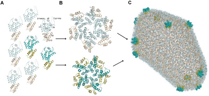
Assembly of the HIV-1 capsid. ( A ) Each capsid monomer is shown…
Figure 1
Assembly of the HIV-1 capsid. (A) Each capsid monomer is shown in cartoon representation, with CA-NTDs in light or dark cyan and CA-CTDs in wheat or olive. Light and dark shades represent monomers that contribute to capsid hexamers and pentamers, respectively. For illustration, on one monomer, α-helices are labelled sequentially, and the positions of N-terminal β-hairpin and Cyp-loop are indicated. (B) Capsid monomers pack into either hexamers (top) or pentamers (bottom). (C) The HIV-1 capsid (PDB: 3J3Q). Approximately 200–250 capsid hexamers combine with 12 capsid pentamers to form a closed fullerene cone structure. Hexamers are shown in cartoon representation. The 12 pentamers are distributed toward the ends of the structure and shown in surface representation.
Figure 2
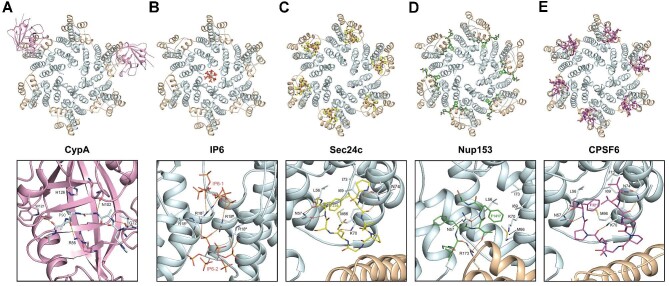
The HIV-1 capsid–natural ligand complexes.…
Figure 2
The HIV-1 capsid–natural ligand complexes. The HIV-1 capsid hexamer bound with the cellular…
Figure 2
The HIV-1 capsid–natural ligand complexes. The HIV-1 capsid hexamer bound with the cellular factor CypA (PDB: 5FJB; A), IP6 (PDB: 6BHT; B), Sec24C (PDB: 8CL3; C), Nup153 (PDB: 6AYA; D), or CPSF6 (PDB: 7ZUD; E). In each panel, the protein backbone of the six capsid protomers is shown in cartoon representation, with CA-NTDs in light cyan and CA-CTDs in wheat. (A) Upper panel: two molecules of CypA are shown in pink cartoon representation bound to Cyp-loops of non-adjacent capsid protomers within the hexamer. Lower panel: the backbone of CypA is shown in a pink cartoon. Capsid residues in the capsid Cyp-loop that are bound in the active site and CypA residues that make contacts are shown as sticks with hydrogen bonds represented by orange dashed lines. (B–E) IP6 (B), the peptides from Sec24C (C), Nup153 (D), and CPSF6 (E) are shown in stick representation. IP6 is coloured by atom type. Sec24C, Nup153, and CPSF6 are coloured in yellow, green, and magenta, respectively. Lower panels: the capsid residues that interact with bound molecules are labelled and shown in sticks with hydrogen bonds represented by orange dashed lines.
Figure 3
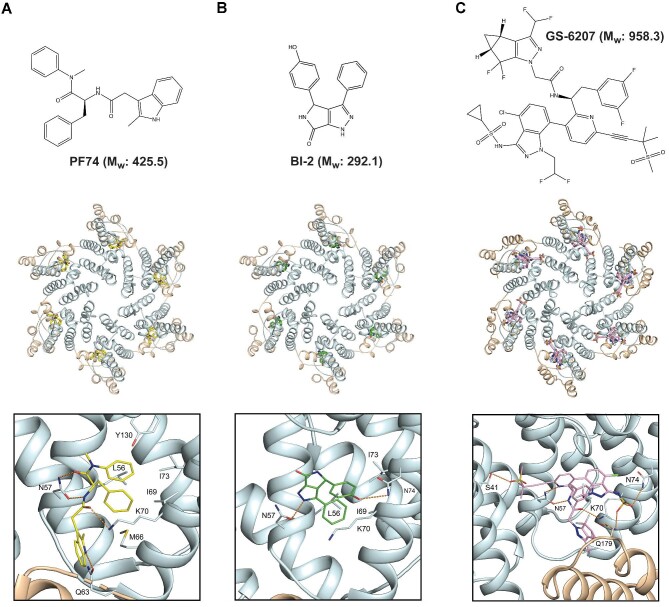
The HIV-1 capsid–drug interactions. Upper…
Figure 3
The HIV-1 capsid–drug interactions. Upper panels: chemical structures and relative molecular weights (M …
Figure 3
The HIV-1 capsid–drug interactions. Upper panels: chemical structures and relative molecular weights (Mw) of drug molecules PF74 (A), BI-2 (B), and GS-6207 (C). Middle panels: the HIV-1 capsid hexamer can bind to PF74 (PDB: 4U0E; A), BI-2 (PDB: 4U0F; B), and GS-6207 (PDB: 6VKV; C). The capsid protein backbone is shown in cartoon representation, with CA-NTDs in light cyan and CA-CTDs in wheat. Drugs are shown in stick representation bound at the α3–α4–α7 pocket. Lower panels: details of molecular interactions at the drug binding sites. Drug molecules and capsid residues that make interactions are shown in stick representation. Hydrogen bonds are represented with orange dashed lines.
Achuthan V., Perreira J.M., Sowd G.A. et al. (2018). Capsid–CPSF6 interaction licenses nuclear HIV-1 trafficking to sites of viral DNA integration. Cell Host Microbe 24, 392–404.
-
PMC
-
PubMed
AlBurtamani N., Paul A., Fassati A. (2021). The role of capsid in the early steps of HIV-1 infection: new insights into the core of the matter. Viruses 13, 1161.
-
PMC
-
PubMed
Ao Z., Danappa Jayappa K., Wang B. et al. (2010). Importin α3 interacts with HIV-1 integrase and contributes to HIV-1 nuclear import and replication. J. Virol. 84, 8650–8663.
-
PMC
-
PubMed
Ay S., Di Nunzio F. (2023). HIV-induced CPSF6 condensates. J. Mol. Biol. 435, 168094.
-
PubMed
Barre-Sinoussi F., Chermann J.C., Rey F. et al. (1983). Isolation of a T-lymphotropic retrovirus from a patient at risk for acquired immune deficiency syndrome (AIDS). Science 220, 868–871.
-
PubMed