. 2023 Nov 15;1(11):1836-1846.
doi: 10.1021/acsaom.3c00312. eCollection 2023 Nov 24.

Waveguide-Integrated Colloidal Nanocrystal Supraparticle Lasers

Affiliations

Waveguide-Integrated Colloidal Nanocrystal Supraparticle Lasers

Pedro Urbano Alves et al. ACS Appl Opt Mater. .

Abstract

Supraparticle (SP) microlasers fabricated by the self-assembly of colloidal nanocrystals have great potential as coherent optical sources for integrated photonics. However, their deterministic placement for integration with other photonic elements remains an unsolved challenge. In this work, we demonstrate the manipulation and printing of individual SP microlasers, laying the foundation for their use in more complex photonic integrated circuits. We fabricate CdSxSe1-x/ZnS colloidal quantum dot (CQD) SPs with diameters from 4 to 20 μm and Q-factors of approximately 300 via an oil-in-water self-assembly process. Under a subnanosecond-pulse optical excitation at 532 nm, the laser threshold is reached at an average number of excitons per CQD of 2.6, with modes oscillating between 625 and 655 nm. Microtransfer printing is used to pick up individual CQD SPs from an initial substrate and move them to a different one without affecting their capability for lasing. As a proof of concept, a CQD SP is printed on the side of an SU-8 waveguide, and its modes are successfully coupled to the waveguide.


Conflict of interest statement

The authors declare no competing financial interest.

Figures

Figure 1
Figure 1
Illustration of the nucleation process occurring inside the emulsion droplets that leads to SPs (a); emission intensity versus pump energy, with laser threshold at approximately 7 nJ, (b) and emission spectra (c) of an SP with a diameter of 9.8 ± 0.5 μm; micrographs of an SP under optical pumping (λpump = 532 nm; see the Optical Characterization Section) below and above the lasing threshold (d, e). The full optical setup can be seen in Figure S3.
Figure 2
Figure 2
SEM image of an SP of approximately 14 μm in diameter (a) and its CL spectrum (b) and discrete Fourier transform analysis (c). The Q-factor estimated from the WGMs was 295 ± 15.
Figure 3
Figure 3
Study on the free parameter b (from Table S3) as a function of the SP radius (a), extracted using eqs 3–5. The black data points correspond to the SPs that did not achieve lasing, and the red data points to those that did. The two data sets are visibly separable (dashed line), suggesting that the capability of an SP of a given size to operate as a laser is strongly intertwined with the parameter b. The average number of excitons ⟨N⟩ at a laser threshold was then calculated for the lasing SPs (b) based on the fitting parameters (eq 2 and Table S3). The optical pump energies required to reach the laser threshold were extracted from Table S2 and included in the callouts. SPs were optically pumped at λpump = 532 nm (see the Optical Characterization Section). The full optical setup can be seen in Figure S3.
Figure 4
Figure 4
Illustration of the transfer printing process applied to the waveguide coupling of an SP: selection of the SP (a); pick up (b, c); selection of the target destination, e.g., substrate with a waveguide (d); and drop off (e, f). Proof of concept with 15 SPs transfer-printed onto a PDMS substrate to mimic the University of Strathclyde logo under white light (g) and a UV light lamp, λlamp = 365 nm (h). Logo used with permission from University of Strathclyde, Glasgow.
Figure 5
Figure 5
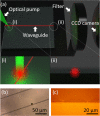
Illustration of the SP-waveguide coupling setup (a), where the sample is simultaneously aligned with the laser pump (a-i) and the CCD camera (a-ii). The SP (diameter ≈7.7 ± 0.5 μm) is being pumped on one edge of the waveguide, and the other edge the facet is being monitored by the CCD camera, which is preceded by a long pass filter (550 nm) to cut out any scattered light from the pump (λpump = 532 nm; see the Optical Characterization Section). The acquired microscope (b) and CCD camera (c) views correspond to the illustrations (a-(i, ii)), respectively. The spectrometer and image readings from the CCD camera were acquired simultaneously and compared to verify which modes were coupled to the waveguide. The full optical setup can be seen in Figure S7.
Figure 6
Figure 6
Readings of the CCD camera, with the enhanced facet pictures and corresponding data, depicted alongside the pictures of the transfer-printed SP (7.7 ± 0.5 μm in diameter) and readings on the spectrometer (a). These measurements were done under four different excitation intensities above the laser threshold. Spectrometer readings below a threshold and under three different excitation intensities are also shown in panel (b). The dashed line seen in spectra (a, b) tracks one of the modes of the SP at approximately 640 nm. The data acquired at that wavelength was used to plot the emission intensity versus pump energy (c), and the laser threshold of the transfer-printed SP was found to be at approximately 3 nJ. The spectral range where the lasing peaks were located in the SP (i.e., from 628 to 648 nm) was divided into 5 equal parts of Δλ = 4 nm each. A Pearson correlation test between the counts registered on the CCD camera and the counts registered on the spectrometer was then performed on each of those 5 parts, using the data of the 4 different optical pump energies (d). The two test parameters, r and p, correspond to the Pearson correlation coefficient and p-value, respectively. SPs were optically pumped at λpump = 532 nm (see the Optical Characterization Section). The full optical setup can be seen in Figure S7.

References

    1. Bera D.; Qian L.; Tseng T. K.; Holloway P. H. Quantum Dots and Their Multimodal Applications: A Review. Materials 2010, 3 (4), 2260–2345. 10.3390/ma3042260. - DOI
    1. Klimov V. I. Spectral and Dynamical Properties of Multiexcitons in Semiconductor Nanocrystals. Annu. Rev. Phys. Chem. 2007, 58 (1), 635–673. 10.1146/annurev.physchem.58.032806.104537. - DOI - PubMed
    1. Ghimire S.; Biju V. Relations of Exciton Dynamics in Quantum Dots to Photoluminescence, Lasing, and Energy Harvesting. J. Photochem. Photobiol., C 2018, 34, 137–151. 10.1016/j.jphotochemrev.2018.01.004. - DOI
    1. Klimov V. I. In Nanocrystal quantum dots: from fundamental photophysics to light-emitting diodes and multicolor lasers , International Conference on Ultimate Lithography and Nanodevice Engineering, 2004.
    1. Wang Y.; Sun H. Advances and Prospects of Lasers Developed from Colloidal Semiconductor Nanostructures. Prog. Quantum Electron. 2018, 60, 1–29. 10.1016/j.pquantelec.2018.05.002. - DOI